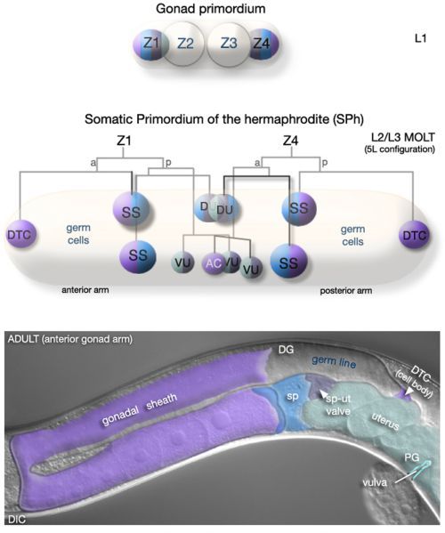
Figure 8. Development of the hermaphrodite somatic gonad.

Figure 8Development of the hermaphrodite somatic gonad

(L1, upper) All cells of the somatic gonad derive from two founder cells, Z1 and Z4, present in the L1 gonad primordium. (Middle-L2/L3 MOLT) By L2, Z1/Z4 have generated 12 descendents: 2 DTCs, required for gonad elongation and germline patterning; 9 blast cells that will, collectively, generate all other adult somatic gonad cells; 1 anchor cell (AC), a transient cell that functions to pattern the cells of the vulva. Somatic and germ cells are intermingled until the L2/L3 molt where upon they rearrange, establishing the general organization of the future gonad. The DTCs are positioned at the anterior and posterior of the developing gonad. The 10 remaining cells gather at the center to form the SPh (Somatic gonadal Primordium of the hermaphrodite), thus dividing the germ line into anterior and posterior populations of cells or arms. (Lower - ADULT) The adult somatic gonad consists of two arms containing five tissues, each with specific functions and distinct anatomical features: the distal tip cells (DTCs), the gonadal sheath, the spermatheca (sp), the spermatheca-uterine (sp-ut) valve, and the uterus. These tissues, in particular the sheath and DTC, are intimately associated with the germ line and play a critical role in its development and its organization and function in the adult. Reprinted with permission from Wormatlas (http://www.wormatlas.org).

From: Hermaphrodite cell-fate specification

Cover of WormBook
WormBook: The Online Review of C. elegans Biology [Internet].
Pasadena (CA): WormBook; 2005-2018.
Copyright © 2005, WormBook Research Community.

All WormBook content, except where otherwise noted, is licensed under a Creative Commons Attribution License, which permits unrestricted use, distribution, and reproduction in any medium, provided the original work is properly cited.

NCBI Bookshelf. A service of the National Library of Medicine, National Institutes of Health.