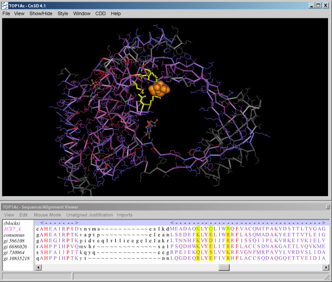
Figure 7. Sequence and structure views of the TOP1Ac conserved domain common to type III bacterial and eukaryotic DNA topoisomerases.

Figure 7Sequence and structure views of the TOP1Ac conserved domain common to type III bacterial and eukaryotic DNA topoisomerases

The upper window displays the structure of the domain with the residues colored according to their sequence conservation, with red indicating high conservation and blue indicating low conservation. The nucleotide bound at site II is shown as an orange space-filling model, and the residues involved in this binding site are yellow. The lower window displays the sequence alignment for the domain with aligned residues shown as colored capital letters. Residues aligned to three of the binding site residues are highlighted in yellow. The sequence for NP_004609 (gi 10835218) occupies the bottom row.

From: Chapter 3, Macromolecular Structure Databases

Cover of The NCBI Handbook
The NCBI Handbook [Internet].
McEntyre J, Ostell J, editors.

NCBI Bookshelf. A service of the National Library of Medicine, National Institutes of Health.