U.S. flag

An official website of the United States government

NCBI Bookshelf. A service of the National Library of Medicine, National Institutes of Health.

Adam MP, Feldman J, Mirzaa GM, et al., editors. GeneReviews® [Internet]. Seattle (WA): University of Washington, Seattle; 1993-2024.

Cover of GeneReviews®

GeneReviews® [Internet].

Show details

Cornelia de Lange Syndrome

Synonyms: BDLS, Brachmann-de Lange Syndrome, CdLS, de Lange Syndrome

, MD, PhD, , MS, and , MD.

Author Information and Affiliations

Initial Posting: ; Last Update: October 15, 2020.

Estimated reading time: 40 minutes

Summary

Clinical characteristics.

Cornelia de Lange syndrome (CdLS) encompasses a spectrum of findings from mild to severe. Severe (classic) CdLS is characterized by distinctive facial features, growth restriction (prenatal onset; <5th centile throughout life), hypertrichosis, and upper-limb reduction defects that range from subtle phalangeal abnormalities to oligodactyly (missing digits). Craniofacial features include synophrys, highly arched and/or thick eyebrows, long eyelashes, short nasal bridge with anteverted nares, small widely spaced teeth, and microcephaly. Individuals with a milder phenotype have less severe growth, cognitive, and limb involvement, but often have facial features consistent with CdLS. Across the CdLS spectrum IQ ranges from below 30 to 102 (mean: 53). Many individuals demonstrate autistic and self-destructive tendencies. Other frequent findings include cardiac septal defects, gastrointestinal dysfunction, hearing loss, myopia, and cryptorchidism or hypoplastic genitalia.

Diagnosis/testing.

The diagnosis of CdLS is established in a proband with suggestive clinical features and/or by identification of a heterozygous pathogenic variant in NIPBL, RAD21, SMC3, or BRD4, or a hemizyous pathogenic variant in HDAC8 or SMC1A by molecular genetic testing.

Management.

Treatment of manifestations: Aggressive management of gastroesophageal reflux with assessment of potential gastrointestinal malrotation; consideration of fundoplication if reflux is severe. Supplementary formulas and/or gastrostomy tube placement to meet nutritional needs as necessary. Physical, occupational, and speech therapy to optimize psychomotor development and communication skills. Standard treatment for epilepsy, vision issues, nasolacrimal duct obstruction, hearing loss, cleft palate, anomalies of tooth formation and/or position, cardiac defects, cryptorchidism/hypospadias, bicornuate uterus, vesicoureteral reflux, anemia and/or thrombocytopenia, and immunodeficiency. If surgery is being considered, malignant hyperthermia precautions and preoperative evaluation for thrombocytopenia and cardiac disease with careful monitoring of the airway during anesthesia are recommended.

Surveillance: At each visit: measurement of growth parameters and evaluation of nutritional status and safety of oral intake; monitor for signs and symptoms of GERD and for evidence of aspiration with respiratory insufficiency; assessment for new manifestations such as seizures or signs of autonomic dysfunction; monitor developmental progress and educational needs; behavioral assessment for anxiety, attention, and aggressive or self-injurious behavior; assessment of mobility and self-help skills. At least annually: ophthalmology evaluation; dental evaluation with cleaning; audiology evaluation in childhood and adolescence.

Genetic counseling.

NIPBL-CdLS, RAD21-CdLS, SMC3-CdLS and BRD4-CdLS are inherited in an autosomal dominant manner; HDAC8-CdLS and SMC1A-CdLS are inherited in an X-linked manner. The majority of affected individuals have a de novo heterozygous pathogenic variant in NIPBL. Fewer than 1% of individuals with autosomal dominant CdLS have an affected parent. When the parents are clinically unaffected, the risk to the sibs of a proband with CdLS is estimated to be 1.5% because of the possibility of germline mosaicism. The risk to sibs of a male proband with X-linked CdLS depends on the status of the proband's mother; the risk to sibs of a female proband with X-linked CdLS depends on the status of the proband's mother and father. Prenatal testing for a pregnancy at increased risk and preimplantation genetic testing are possible for families in which the pathogenic variant has been identified.

Diagnosis

Cornelia de Lange syndrome (CdLS) constitutes a clinical spectrum, with some individuals having milder features and others displaying more severe, classic features. An international consensus statement has defined both cardinal and suggestive features, as well as a scoring system to define classic and non-classic CdLS to assist with clinical genetic testing decisions [Kline et al 2018].

Suggestive Findings

Cornelia de Lange syndrome (CdLS) should be suspected in individuals with the following clinical and radiographic features.

Clinical findings

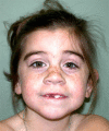
  • Distinctive craniofacial appearance (often recognizable; see Figure 1) including:
    • Microcephaly (mean occipital frontal circumference <2nd centile)
    • Synophrys with highly arched and/or thick eyebrows
    • Long, thick eyelashes
    • Short nasal bridge, upturned nasal tip with anteverted nares
    • Long and/or smooth philtrum, thin vermilion of the upper lip, downturned corners of the mouth
    • Highly arched palate with or without cleft palate
    • Small widely spaced teeth
    • Micrognathia with or without mandibular spurs
  • Growth failure, which may be detected prenatally and persists postnatally
  • Developmental delay / intellectual disability, varying from mild to profound
  • Limb abnormalities (either symmetric or asymmetric) ranging from severe reduction defects with complete absence of the forearms, to various forms of oligodactyly (missing digits) involving primarily the upper extremities, to small hands (micromelia) and phalangeal abnormalities (5th digit clinodactyly and short first metacarpal resulting in a proximally placed thumb) at the mild end
  • Hypertrichosis. Thick scalp hair that often extends onto the temporal regions and at times involves the face, ears, back, and arms
Figure 1.

Figure 1.

Classic CdLS craniofacial features

Radiographic findings. In those without limb deficiencies, the presence of a short first metacarpal on plain x-ray resulting in a proximally placed thumb can be useful in diagnosis.

Establishing the Diagnosis

The diagnosis of CdLS is established in a proband with the above clinical features and/or by the identification on molecular genetic testing of a heterozygous pathogenic (or likely pathogenic) variant in NIPBL, RAD21, SMC3, or BRD4 or a hemizyous pathogenic (or likely pathogenic) variant in HDAC8 or SMC1A (see Table 1).

Note: (1) Per ACMG/AMP variant interpretation guidelines, the terms "pathogenic variants" and "likely pathogenic variants" are synonymous in a clinical setting, meaning that both are considered diagnostic and both can be used for clinical decision making [Richards et al 2015]. Reference to "pathogenic variants" in this section is understood to include any likely pathogenic variants. (2) The identification of variant(s) of uncertain significance cannot be used to confirm or rule out the diagnosis.

Molecular genetic testing approaches can include a combination of gene-targeted testing (serial single-gene testing or multigene panel) and comprehensive genomic testing (exome sequencing, exome array, genome sequencing) depending on the phenotype.

Gene-targeted testing requires that the clinician determine which gene(s) are likely involved, whereas genomic testing does not. Because the phenotype of Cornelia de Lange syndrome can be broad, individuals with distinctive features described in Suggestive Findings are likely to be diagnosed using gene-targeted testing (see Option 1), whereas those in whom the diagnosis of Cornelia de Lange syndrome has not been considered are more likely to be diagnosed using genomic testing (see Option 2).

Option 1

When the phenotypic findings suggest the diagnosis of Cornelia de Lange syndrome, molecular genetic testing approaches include use of a multigene panel or serial single gene testing.

A Cornelia de Lange syndrome multigene panel that contains at least NIPBL, SMC1A, HDAC8, SMC3, RAD21 and BRD4 and several additional genes that can cause a phenotype resembling CdLS, such as AFF4, ANKRD11, CREBBP, and EP300 (see Differential Diagnosis), is the most effective way of detecting causal variants [Kline et al 2018]. Because of the significant frequency of somatic mosaicism [Huisman et al 2013], testing capable of detecting mosaicism, such as next generation sequencing (NGS), should be considered, preferably using uncultured fibroblasts, although buccal cells or bladder epithelial cells can also be used.

This approach is most likely to identify the genetic cause of the condition while limiting identification of variants of uncertain significance and pathogenic variants in genes that do not explain the underlying phenotype. Note: (1) The genes included in the panel and the diagnostic sensitivity of the testing used for each gene vary by laboratory and are likely to change over time. (2) Some multigene panels may include genes not associated with the condition discussed in this GeneReview. (3) In some laboratories, panel options may include a custom laboratory-designed panel and/or custom phenotype-focused exome analysis that includes genes specified by the clinician. (4) Methods used in a panel may include sequence analysis, deletion/duplication analysis, and/or other non-sequencing-based tests. For CdLS, a multigene panel that also includes deletion/duplication analysis is recommended (see Table 1).

For an introduction to multigene panels click here. More detailed information for clinicians ordering genetic tests can be found here.

Serial single-gene testing. Sequential molecular genetic testing of NIPBL, SMC1A, HDAC8, SMC3, RAD21 and BRD4 may be considered:

  • For individuals with features strongly suggestive of CdLS who score highly on the international consensus scoring criteria or for whom multigene panel testing is not available, NIPBL sequence analysis may be considered first. If no pathogenic variant is identified, gene-targeted deletion/duplication analysis of NIPBL should be considered next.
  • If no NIPBL pathogenic variant is identified and the affected individual has milder physical features of CdLS, consider SMC1A sequence and gene-targeted deletion/duplication analysis next.
  • If no NIPBL or SMC1A pathogenic variant is identified and CdLS is highly suspected (especially in an individual with milder features), consider BRD4, SMC3, RAD21, and HDAC8 sequence analysis and gene-targeted deletion/duplication analysis.

Option 2

When the diagnosis of Cornelia de Lange syndrome is not strongly considered due to atypical phenotypic features, comprehensive genomic testing (which does not require the clinician to determine which gene[s] are likely involved) is the best option. Exome sequencing is most commonly used; genome sequencing is also possible.

For an introduction to comprehensive genomic testing click here. More detailed information for clinicians ordering genomic testing can be found here.

Table 1.

Molecular Genetic Testing Used in Cornelia de Lange Syndrome

Gene 1, 2Proportion of CdLS
Attributed to Pathogenic
Variants in Gene
Proportion of Probands with a Pathogenic Variant 3 Detectable by Method
Sequence
analysis 4
Gene-targeted deletion/duplication analysis 5
BRD4 <1%3/4 61/4 6
HDAC8 ~4%~90% 7~10% 7
NIPBL ~80% 8, 9~97% 9~3% 10, 11
RAD21 <1% 1220/22 122/22 12
SMC1A ~5% 13~100%
SMC3 1%-2% 14~97%~3%
Unknown3%-5%
1.

Genes are listed in alphabetic order.

2.

See Table A. Genes and Databases for chromosome locus and protein.

3.

See Molecular Genetics for information on allelic variants detected in these genes.

4.

Sequence analysis detects variants that are benign, likely benign, of uncertain significance, likely pathogenic, or pathogenic. Variants may include small intragenic deletions/insertions and missense, nonsense, and splice site variants; typically, exon or whole-gene deletions/duplications are not detected. For issues to consider in interpretation of sequence analysis results, click here.

5.

Gene-targeted deletion/duplication analysis detects intragenic deletions or duplications. Methods used may include a range of techniques such as quantitative PCR, long-range PCR, multiplex ligation-dependent probe amplification (MLPA), and a gene-targeted microarray designed to detect single-exon deletions or duplications. Gene-targeted deletion/duplication testing will detect deletions ranging from a single exon to the whole gene; however, breakpoints of large deletions and/or deletion of adjacent genes (e.g., those described by Olley et al [2018]) may not be detected by these methods.

6.
7.
8.
9.

Somatic mosaicism for NIPBL has been reported in approximately 10%-15% of individuals. Obtaining a buccal sample at the time of collecting a peripheral blood sample can be considered [Huisman et al 2013]. Screening for NIPBL mosaicism can be pursued if CdLS is strongly suspected and molecular genetic testing on a peripheral blood sample is normal.

10.

Gene-targeted deletion/duplication analysis of NIPBL detects ~3% of NIPBL-CdLS [Pehlivan et al 2012, Russo et al 2012, Ansari et al 2014].

11.

Cytogenetic testing or chromosomal microarray (CMA) may also be considered in those with classic features of CdLS but normal molecular genetic testing because a few individuals with large deletions of 5p13 that include NIPBL have been reported [Taylor & Josifek 1981, Hulinsky et al 2005, Hayashi et al 2007].

12.
13.
14.

Clinical Characteristics

Clinical Description

While classic Cornelia de Lange syndrome (CdLS) was formally characterized more than 70 years ago and well delineated clinically [Ptacek et al 1963, Jackson et al 1993], the identification of the molecular genetic basis of CdLS has led to the recognition of affected individuals who have milder or atypical features. Therefore, this condition encompasses a spectrum of findings from mild to severe (see Phenotype Correlations by Gene). Those with a milder phenotype, which is less striking clinically than the classic form of CdLS, may represent the majority of individuals with CdLS [Deardorff et al 2007, Rohatgi et al 2010].

Table 2.

Features of Cornelia de Lange Syndrome

Feature% of Persons w/FeatureComment
Synophrys98% 1In individuals w/classic features
Feeding difficulties>95%
Growth failure>95%May be noted prenatally
Intellectual disability>95%Typically severe to profound in those w/classic CdLS
Small hands & feet>90%
Microbrachycephaly>90%
Long eyelashes>90%
Thin upper vermilion border of lip>90%
Downturned corners of mouth>90%
Dental problems>90%Delayed secondary tooth eruption, small or absent teeth, malposition, overcrowding, caries due to GERD, periodontal disease, & bruxism
Hypertrichosis>80%Involving the face, ears, back, & arms
Hearing loss80%>40% have profound SNHL, although hearing loss may improve over time.
Micrognathia80%In those w/classic features
Radial head underdevelopment79%
Gastroesophageal reflux disease75%
Clinodactyly70%
Nasolacrimal duct obstruction70%
Ptosis60%
Cutis marmorata60%
Self-injurious behavior56%
Sleep difficulties50%
Mandibular spurs42%
Scoliosis33%
High & arched palate w/clefting30%
Congenital heart defects30%Most commonly pulmonic or peripheral pulmonic stenosis, VSD, & ASD
Seizures25%
Oligodactyly25%

Features and frequencies are largely derived from Jackson et al [1993], Kline at al [2018] and references therein.

ASD = atrial septal defect; GERD = gastroesophageal disease; SNHL = sensorineural hearing loss; VSD = ventricular septal defect

Growth. Prenatal-onset growth failure is present in a majority of individuals with CdLS. Symmetric slow growth resulting in proportionate short stature becomes more significant by age six months. Mean height and weight are below the fifth centile throughout life [Boog et al 1999] in individuals with classic CdLS. Growth may be less severely affected in those with overall milder clinical features and/or mosaic pathogenic variants. In addition, failure to thrive may be superimposed on the constitutional growth restriction secondary to gastroesophageal reflux disease and other issues with feeding (see Gastrointestinal below).

CdLS-specific growth charts have been developed. See www.cdlsusa.org (girls; boys).

Intellectual disability. Most individuals with CdLS have developmental delay. The overall range of IQ levels is broad, from below 30 to 102, with an average IQ of 53 [Kline et al 1993, Saal et al 1993]. Those affected individuals with classic features are more likely to have severe-to-profound intellectual disability (see Genotype-Phenotype Correlations).

  • Expressive language is often more compromised than receptive language, and receptive language more compromised than cognition [Ajmone et al 2014].
  • Effective verbal and nonverbal communication skills can facilitate quality of life enormously [Kline et al 2018]. Early augmentative and alternative communication interventions are highly effective [Ajmone et al 2014].
  • Half of children walk by 24 months and 95% by age ten. Half of children are able to feed themselves by age three years and 95% by age ten [Kline et al 1993].

Behavior. A range of behavioral issues have been reported [Kline et al 2018].

  • Behavior problems are often directly related to frustration from an inability to communicate (see Intellectual disability above).
  • Difficulties in sensory processing can lead to hyposensitivity, hypersensitivity, disorientation, and fixation.
  • Autistic behavior may lead to avoidance or rejection of social interaction and physical contact.
  • Repetitive behaviors, which can be exacerbated by anxiety, are common and are associated with more marked intellectual disability and autistic features.
  • Clinically significant self-injurious behavior occurs in 56% of individuals, with hand-directed self-injurious behavior the most common.

Neurologic. Overall, approximately 25% of individuals with CdLS have seizures. Partial epilepsy is the most common type with age of onset typically before age two years. Most affected individuals respond well to standard medical therapy and are able to be weaned off medical therapy after a few years [Verrotti et al 2013] (see Management, Treatment of Manifestations).

Limb involvement. Severe abnormalities of the upper extremities are seen in 25% of individuals with CdLS.

  • Upper-extremity deficiencies ranging from severe reduction defects with complete absence of the forearms to various forms of oligodactyly (missing digits) are present in about one third of those with classic features.
  • In the absence of limb deficiency, micromelia (small hands), proximally placed thumbs, and fifth-finger clinodactyly occur in nearly all individuals (see Figure 3).
  • Radioulnar synostosis is common and may result in flexion contractures of the elbows.
  • The lower extremities are less involved than the upper extremities. The feet are often small and syndactyly of the second and third toes occurs in more than 80% of affected individuals [Jackson et al 1993].
Figure 3.

Figure 3.

Range of limb anomalies in CdLS

Gastrointestinal. Gastroesophageal reflux disease (GERD) is present in a majority of affected indivdiuals. Other complications of GERD including esophagitis, aspiration, chemical pneumonitis, and irritability can be avoided by diagnosis and treatment of GERD in the neonatal period (see Management). Approximately one third have evidence of aspiration and 15% require a feeding tube [Luzzani et al 2003]. Other gastrointestinal abnormalities include:

  • Pyloric stenosis (4%), the most frequent cause of persistent vomiting in the newborn period
  • Intestinal malrotation (2%)
  • CDH has been diagnosed both pre- and postnatally, but may be underascertained, especially in infants who die in the perinatal period.

Ophthalmologic. As many as 60% of affected individuals demonstrate some degree of ptosis as well as other ocular problems including myopia (60%) and nystagmus (37%) [Levin et al 1990]. Other ophthalmologic abnormalities:

  • Nasolacrimal duct stenosis
  • Glaucoma
  • Microcornea
  • Astigmatism
  • Optic atrophy
  • Coloboma of the optic nerve
  • Strabismus
  • Proptosis

Otolaryngologic. Sensorineural hearing impairment is noted in 40% of children with CdLS, and conductive hearing impairment in 60% [Marchisio et al 2014]. Notably, for a significant proportion hearing loss (both sensorineural and conductive) improves over time [Janek et al 2016]

Genitourinary. Renal abnormalities, primarily vesicoureteral reflux, have been reported in 12%.

  • Cryptorchidism occurs in 73% of males with CdLS
  • Hypoplastic (small) genitalia occur in 57% and hypospadias in 9% of males [Jackson et al 1993]
  • Bicornuate uterus, which can cause abdominal pain, has been observed in five (25%) of 20 affected females [Oliver et al 2010, Kline et al 2015].

Cardiovascular. Approximately 30% of individuals with CdLS have congenital heart disease [Chatfield et al 2012]. The most common abnormalities include (in descending order):

  • Pulmonic or peripheral pulmonic stenosis
  • Ventricular septal defects
  • Atrial septal defects
  • Coarctation or hypoplastic aortic arch
  • Aortic valve anomaly
  • Tetralogy of Fallot
  • Double-outlet right ventricle
  • Atrioventricular canal defect

Immunologic. Antibody deficiency has been described in several individuals with CdLS, indicating a need for immunologic screening and management of immunodeficiency in those affected individuals with severe or recurrent infections [Jyonouchi et al 2013]. The most common reported recurrent infections include chronic ear infections, chronic viral respiratory infections, and pneumonia. Impaired T-cell function may be associated with antibody deficiencies observed in persons with CdLS.

Hematologic. Thrombocytopenia often resolves after infancy, but can rarely transition to persistent idiopathic thrombocytopenia purpura [Lambert et al 2011]. In most cases, anemia is transient.

Other features

  • A characteristic low-pitched cry that tends to disappear in late infancy has been described in 75% of children with CdLS and is associated with more severe cases [Jackson et al 1993].
  • Hypoplastic nipples and umbilicus are seen in 50%.

Prognosis. The majority of familial cases suggest that expressivity is relatively consistent within a family. In the absence of severe congenital anomalies of internal organs, life expectancy is not significantly reduced.

  • Beck & Fenger [1985] studied mortality in 48 individuals with CdLS born between 1917 and 1982 and found a modest increase in mortality over the general population when comparing cumulative survival rates; the increase is more significant among the younger age groups. They also reported two individuals who died at ages 54 and 61 years.
  • Schrier et al [2011] reported 295 affected individuals in whom a cause of death was known. Respiratory causes, including aspiration/reflux and pneumonias, were the most common primary causes (31%), followed by gastrointestinal disease, including obstruction/volvulus (19%). Congenital anomalies accounted for 15% of deaths and included congenital diaphragmatic hernia and congenital heart defects. Acquired cardiac disease accounted for 3% of deaths. Neurologic causes and accidents each accounted for 8%, sepsis for 4%, cancer for 2%, renal disease for 1.7%, and other causes 9% of deaths.

Phenotype Correlations by Gene

NIPBL

Individuals with classic findings of CdLS, including characteristic facial features and limb anomalies, are more likely to have a pathogenic variant in NIPBL.

Other Genes

Milder phenotypes that retain some of the characteristic facial features but with variable cognitive and limb or structural involvement compared to individuals with a pathogenic variant in NIPBL have been consistently described and can be seen in individuals with a heterozygous pathogenic variant in BRD4, SMC3 or RAD21 or a hemizygous pathogenic variant in HDAC8 or SMC1A [Deardorff et al 2007, Selicorni et al 2007, Rohatgi et al 2010, Kaiser et al 2014, Minor et al 2014, Gil-Rodríguez et al 2015, Olley et al 2018] (see Figure 2).

Figure 2.

Figure 2.

Affected individual with a pathogenic variant in SMC1A

SMC1A and SMC3

  • Individuals with SMC1A or SMC3 pathogenic variants typically have fewer structural anomalies and less severe growth restriction than those with NIPBL pathogenic variants; however, they have significant intellectual disability that can range from moderate to severe [Deardorff et al 2007, Gil-Rodríguez et al 2015, Huisman et al 2017].
  • Compared to individuals with a heterozygous NIPBL pathogenic variant, the facial features in those with SMC1A or SMC3 pathogenic variants include slightly flatter and broader eyebrows and a broader and longer nasal bridge [Rohatgi et al 2010].
  • Those with pathogenic variants in SMC3 specifically often have subtle or absent synophrys, wider bulbous nose, and a long but well-formed philtrum.
  • Cardiac malformations, although typically mild, are also observed (~56%) in individuals with SMC3 pathogenic variants, and less frequently in individuals with SMC1A pathogenic variants [Gil-Rodríguez et al 2015].

RAD21. Individuals with a heterozygous RAD21 pathogenic variant:

  • Typically do not have major structural differences.
  • Have milder cognitive impairment compared to those with classic CdLS.
    Specifically, 10% have normal cognition, 45% have mild cognitive impairment and none have severe or profound cognitive disability [Krab et al 2020].
  • Often display growth restriction, minor skeletal anomalies, and facial features that overlap with CdLS [Deardorff et al 2012, Krab et al 2020].

HDAC8

  • Males with a hemizygous pathogenic variant in HDAC8 have facial features that overlap with CdLS but typically display delayed closure of the anterior fontanelle, hooded eyelids, widely spaced eyes, a wide nose, mosaic skin pigmentation, dental anomalies, and happy or friendly personalities. Growth also tends to be less severely affected with lower frequency of postnatal growth restriction and microcephaly reported.
  • Females. The clinical presentation for a female with a heterozygous pathogenic variant in HDAC8 overlaps that of males, but the severity is greatly influenced by the pattern of X inactivation [Kaiser et al 2014].

Genotype-Phenotype Correlations

NIPBL. Individuals with pathogenic missense variants and in-frame deletions in NIPBL have been found to have less severe growth deficiency, milder intellectual disability, and fewer structural anomalies [Gillis et al 2004, Ajmone et al 2014, Ansari et al 2014] compared to those with pathogenic loss-of-function NIPBL variants.

SMC1A. Pathogenic variants that cause a CdLS phenotype are typically missense and can result in a range of severity. Of note, loss of function pathogenic variants in SMC1A cause an early-infantile epileptic encephalopathy (OMIM 301044; see Genetically Related Disorders).

Nomenclature

Cornelia de Lange syndrome (CdLS) was first described by Vrolik in 1849, who reported a case as an extreme example of oligodactyly [Oostra et al 1994]. Brachmann [1916] provided a detailed account of a case of symmetric monodactyly, antecubital webbing, dwarfism, cervical ribs, and hirsutism.

In the 1930s, Cornelia de Lange, a Dutch pediatrician, described two unrelated girls with similar features and named the condition after the city in which she worked: typus degenerativus amstelodamensis [de Lange 1933, de Knecht-van Eekelen & Hennekam 1994]. Some examples in the literature refer to the disorder as Brachmann-de Lange syndrome; however, it is more widely referred to as Cornelia de Lange syndrome in honor of Dr de Lange’s contributions to the understanding of the disorder.

Prevalence

The prevalence of CdLS is difficult to estimate as individuals with milder or variable features are likely under-recognized. Published estimates for the prevalence range from 1:100,000 [Pearce & Pitt 1967] to as high as 1:10,000 [Opitz 1985]. Data from the EUROCAT dataset have estimated the prevalence at 1:50,000 for the classic form of CdLS [Barisic et al 2008]; this figure is less likely to include the milder, more common phenotype.

Differential Diagnosis

Phenotypic overlap with CdLS may be observed in monogenic disorders (see Table 4), chromosome abnormalities, and fetal alcohol syndrome.

Table 4.

Genes of Interest in the Differential Diagnosis of Cornelia de Lange Syndrome

GeneDisorderMOIFeatures of Differential Disorder
Overlapping w/CdLSDistinguishing from CdLS
AFF4 CHOPS syndrome (OMIM 616368)ADCognitive impairment, heart defects, short stature, skeletal dysplasiaCoarse facial features, obesity, & pulmonary involvement (features that are not typical for CdLS) 1
ANKRD11 2 KBG syndrome ADDD/ID, short stature, thick eyebrows & synophrys, anteverted nares 3ID is typically milder; macrodontia, few congenital defects
ASXL1 Bohring-Opitz syndrome (BOS)ADDD/ID; prenatal & postnatal growth restriction, microcephaly, hypertrichosis, small feet, facial resemblance w/CdLS (especially infants)Feeding intolerance is usually more severe; high myopia, BOS posture 4, prominent globes
EP300 EP300 Rubinstein-Taybi Syndrome (RSTS)ADDD/ID, small stature, hypertrichosis, full eyebrows, long lashes, micrognathia, malrotation 5Prominent nose & broad thumbs (both less common in EP300-RSTS than in CREBBP-RSTS) 6
TAF1 Intellectual disability syndrome (OMIM 300966)XLDD/ID, long philtrum, anteverted nares, microcephaly, hearing loss 7Generalized hypotonia, long face, protruding ears
TAF6 Alazami-Yuan syndrome (OMIM 617126)ARShort stature, microcephaly, clinodactyly, hirsutism, DD/ID, & facial dysmorphism w/long philtrum, thin & arched eyebrows, synophrys 8Widow’s peak (rare)

AD = autosomal dominant; AR = autosomal recessive; CdLS = Cornelia de Lange syndrome; DD = developmental delay; ID = intellectual disability; MOI = mode of inheritance; XL = X-linked

1.
2.

KBG syndrome is caused by either a heterozygous pathogenic variant in ANKRD11 or deletion of 16q24.3 that includes ANKRD11.

3.
4.

Individuals with Bohring-Opitz syndrome (BOS) can have a specific limb posture termed "BOS posture," described as an external rotation or adduction of the shoulders with flexion of the wrists and fingers at the metacarpophalangeal joint.

5.
6.

EP300 pathogenic variants cause a phenotype that resembles RSTS caused by mutation of CREBBP. However, with the exception of the low-hanging columella, the facial features in EP300-RSTS are less marked. Although the thumbs and halluces are broad, angulation is very uncommon. Intellectual disability is variable but is usually less severe and occasionally normal (see Rubinstein-Taybi Syndrome).

7.
8.

Deletions of chromosome 2q31.1 (OMIM 613681). Deletions in this region that encompass the HOXD cluster produce limb reduction defects similar to those seen in CdLS as well as genitourinary and developmental abnormalities [Del Campo et al 1999]. Individuals with deletion of 2q31.1 do not have the characteristic facies of CdLS.

Fryns syndrome can be considered in the differential diagnosis of CdLS, most often due to the presence of diaphragmatic defects (diaphragmatic hernia, eventration, hypoplasia, or agenesis) in both disorders. Fryns syndrome and CdLS are characterized by cardiac and renal anomalies, as well as cleft palate, long philtrum, micrognathia, and distal digital hypoplasia (nails, terminal phalanges). However, other features of Fryns syndrome vary (e.g., widely spaced eyes, broad nasal bridge, and macrostomia). While survival beyond the neonatal period has been rare for Fryns syndrome, severe developmental delay and intellectual disability are common. The molecular diagnosis of Fryns syndrome can be established in proband with suggestive findings and biallelic pathogenic variants in PIGN identified by molecular genetic testing. Genetic heterogeneity for Fryns syndrome remains highly probable, as some individuals with a clinical diagnosis of Fryns syndrome have not had PIGN pathogenic variants identified. See also Congenital Diaphragmatic Hernia Overview.

Fetal alcohol syndrome (FAS). Features common to both FAS and CdLS include intrauterine growth restriction, failure to thrive, developmental abnormalities, hyperactivity, microcephaly, facial hypertrichosis in the newborn, short palpebral fissures, short nose with anteverted nares, long and smooth philtrum, thin vermilion of the upper lip, and cardiac defects. However, the hands and feet in FAS are not small and speech is less affected than in CdLS. A history of alcohol use during pregnancy is useful in discriminating FAS from CdLS.

Management

Clinical management guidelines for Cornelia de Lange syndrome have been published [Kline et al 2007, Kline et al 2018] (full text).

Evaluations Following Initial Diagnosis

To establish the extent of disease and needs in an individual diagnosed with Cornelia de Lange syndrome, the evaluations summarized in Table 5 (if not performed as part of the evaluation that led to the diagnosis) are recommended.

Table 5.

Recommended Evaluations Following Initial Diagnosis in Individuals with Cornelia de Lange Syndrome

System/ConcernEvaluationComment
Constitutional Measurement of growth parametersCdLS-specific growth charts are available. 1
Neurologic Neurologic evalConsider EEG if seizures are a concern.
Development Developmental assessment
  • Motor, adaptive, cognitive, & speech/language eval. Speech therapy is highly recommended to optimize communication skills & should be implemented in 1st 18 mos of life.
  • Eval for early intervention / special education
Behavioral Neuropsychiatric evalFor persons age >12 mos: screen for behavior concerns incl sleep disturbances, ADHD, anxiety, &/or findings suggestive of ASD.
Musculoskeletal Consider radiographs of upper extremitiesTo assess for radioulnar synostosis
Orthopedics / physical medicine & rehab / PT & OT evalTo incl assessment of:
  • Gross motor & fine motor skills
  • Limb deficiencies
  • Scoliosis
  • Mobility, activities of daily living, & need for adaptive devices
  • Need for PT (to improve gross motor skills) &/or OT (to improve fine motor skills)
Gastrointestinal/
Feeding
Gastroenterology / nutrition / feeding team eval
  • Consider upper GI series to evaluate for malrotation.
  • Consider endoscopy, pH probe for severe or refractory GERD.
  • Eval of aspiration risk
  • Eval of nutritional status
  • Consider eval for gastric tube placement in those w/dysphagia &/or aspiration risk.
Eyes Ophthalmologic evalIncl eval for nasolacrimal duct patency, ptosis, assessment of visual acuity, dilated fundus exam, measurement of intraocular pressure
Hearing Audiologic eval 2Assess for hearing loss.
ENT/Mouth Clinical assessment for cleft palate
  • Examination of palate by both inspection & palpation at diagnosis
  • In case of symptoms of a (submucous) cleft palate, referral for specialist assessment is indicated.
Dental Dental assessmentIn infancy in those w/cleft palate or after tooth eruption
Cardiovascular EchocardiogramAssessment for congenital heart defects
Genitourinary Assessment for cryptorchidism &/or hypospadias in malesConsider referral to urologist.
Renal ultrasoundEvaluate for structural kidney anomalies.
Consider VCUG.To assess for vesicoureteral reflux, as clinically indicated
Consider pelvic ultrasound.In females of pubertal age to screen for uterine anomalies
Hematologic Complete blood countIf signs of anemia, bruising, &/or bleeding
Immunologic Complete blood count & immune profile 3If recurrent infections are present, consider referral to immunologist.
Miscellaneous/
Other
Consultation w/clinical geneticist &/or genetic counselorTo incl genetic counseling
Family support
& resources
Assess need for:

ASD = autism spectrum disorder; GERD = gastrointestinal reflux disease; OT = occupational therapy; PT = physical therapy; VCUG = vesicoureterogram

1.
2.

To include auditory brain stem response testing and otoacoustic emissions testing

3.

Quantitative immunoglobulins; antibodies to tetanus, diptheria, and pneumococcus; B-cell panel; T-cell panel

Treatment of Manifestations

Table 6.

Treatment of Manifestations in Individuals with Cornelia de Lange Syndrome

Manifestation/
Concern
TreatmentConsiderations/Other
Poor weight gain /
Failure to thrive
Feeding therapy; gastrostomy tube placement may be required for persistent feeding issues.
  • Low threshold for clinical feeding eval &/or radiographic swallowing study if clinical signs or symptoms of dysphagia
  • Referral to a nutritionist may be considered.
Gastroesophageal
reflux disease
  • Proactive mgmt of GERD w/very ↓ threshold for medical therapy
  • Standard medication & postprandial positioning
Consider fundoplication if symptoms are severe.
Malrotation Surgical correction
Epilepsy Standard treatment w/ASM by experienced neurologist
  • Many ASMs may be effective; none has been demonstrated effective specifically for CdLS.
  • Education of parents/caregivers 1
DD/ID See Developmental Delay / Intellectual Disability Management Issues.Early implementation of speech therapy
Limb defects Consideration of surgical intervention of arms/handsRarely needed
Orthopedics / physical medicine & rehab / PT & OTConsider need for positioning & mobility devices, disability parking placard.
Ptosis, strabismus,
&/or abnormal vision
Standard treatment(s) as recommended by ophthalmologistCommunity vision services through early intervention or school district
Nasolacrimal duct
obstruction
Aggressive treatment per ophthalmologistMassage therapy is often unsuccessful because of narrow malformed ducts.
Hearing Hearing aids may be helpful; per otolaryngologist.Community hearing services through early intervention or school district
Cleft palate Standard treatment per multidisciplinary craniofacial team, if possible
Anomalies of tooth
formation &/or
positioning
Standard treatment per dentist &/or orthodontist
Congenital heart
defects
Standard treatment per cardiologist
Cryptorchidism/
Hypospadias
Standard treatment by urologist
Vesicoureteral
reflux
Bicornuate uterus Standard treatment per gynecologist
Anemia &/or
thrombocytopenia
Eval by hematologistSevere thrombocytopenia may require IVIgG &/or steroid treatment.
Immunodeficiency Standard treatment by immunologistUnless specific concerns, routine immunizations should be given.
Surgical risks
  • Preoperative eval for thrombocytopenia & cardiac disease
  • Sedation &/or operative procedures w/anesthesiologists experienced in mgmt of small airways present in CdLS
Adverse reactions to midazolam & malignant hyperthermia have been seen, although rare 2 (see Malignant Hyperthermia Susceptibility).
Family/Community
  • Ensure appropriate social work involvement to connect families w/local resources, respite, & support.
  • Coordinate care to manage multiple subspecialty appointments, equipment, medications, & supplies.
  • Ongoing assessment of need for palliative care involvement &/or home nursing
  • Consider involvement in adaptive sports or Special Olympics.

ASM = anti-seizure medication; DD/ID = developmental delay / intellectual disability; GERD = gastroesophageal reflux disease; OT = occupational therapy; PT = physical therapy

1.

Education of parents/caregivers regarding common seizure presentations is appropriate. For information on non-medical interventions and coping strategies for children diagnosed with epilepsy, see Epilepsy Foundation Toolbox.

2.

Developmental Delay / Intellectual Disability Management Issues

The following information represents typical management recommendations for individuals with developmental delay / intellectual disability in the United States; standard recommendations may vary from country to country.

Ages 0-3 years. Referral to an early intervention program is recommended for access to occupational, physical, speech, and feeding therapy as well as infant mental health services, special educators, and sensory impairment specialists. In the US, early intervention is a federally funded program available in all states that provides in-home services to target individual therapy needs.

Ages 3-5 years. In the US, developmental preschool through the local public school district is recommended. Before placement, an evaluation is made to determine needed services and therapies and an individualized education plan (IEP) is developed for those who qualify based on established motor, language, social, or cognitive delay. The early intervention program typically assists with this transition. Developmental preschool is center based; for children too medically unstable to attend, home-based services are provided.

All ages. Consultation with a developmental pediatrician is recommended to ensure the involvement of appropriate community, state, and educational agencies (US) and to support parents in maximizing quality of life. Some issues to consider:

  • IEP services:
    • An IEP provides specially designed instruction and related services to children who qualify.
    • IEP services will be reviewed annually to determine whether any changes are needed.
    • Special education law requires that children participating in an IEP be in the least restrictive environment feasible at school and included in general education as much as possible, when and where appropriate.
    • Vision and hearing consultants should be a part of the child's IEP team to support access to academic material.
    • PT, OT, and speech services will be provided in the IEP to the extent that the need affects the child's access to academic material. Beyond that, private supportive therapies based on the affected individual's needs may be considered. Specific recommendations regarding type of therapy can be made by a developmental pediatrician.
    • As a child enters the teen years, a transition plan should be discussed and incorporated in the IEP. For those receiving IEP services, the public school district is required to provide services until age 21.
  • A 504 plan (Section 504: a US federal statute that prohibits discrimination based on disability) can be considered for those who require accommodations or modifications such as front-of-class seating, assistive technology devices, classroom scribes, extra time between classes, modified assignments, and enlarged text.
  • Developmental Disabilities Administration (DDA) enrollment is recommended. DDA is a US public agency that provides services and support to qualified individuals. Eligibility differs by state but is typically determined by diagnosis and/or associated cognitive/adaptive disabilities.
  • Families with limited income and resources may also qualify for supplemental security income (SSI) for their child with a disability.

Motor Dysfunction

Gross motor dysfunction

  • Physical therapy is recommended to maximize mobility and to reduce the risk for later-onset orthopedic complications (e.g., contractures, scoliosis, hip dislocation).
  • Consider use of durable medical equipment and positioning devices as needed (e.g., wheelchairs, walkers, bath chairs, orthotics, adaptive strollers).
  • For muscle tone abnormalities including hypertonia or dystonia, consider involving appropriate specialists to aid in management of baclofen, tizanidine, Botox®, anti-parkinsonian medications, or orthopedic procedures.

Fine motor dysfunction. Occupational therapy is recommended for difficulty with fine motor skills that affect adaptive function such as feeding, grooming, dressing, and writing.

Oral motor dysfunction should be assessed at each visit and clinical feeding evaluations and/or radiographic swallowing studies should be obtained for choking/gagging during feeds, poor weight gain, frequent respiratory illnesses, or feeding refusal that is not otherwise explained. Assuming that the child is safe to eat by mouth, feeding therapy (typically from an occupational or speech therapist) is recommended to help improve coordination or sensory-related feeding issues. Feeds can be thickened or chilled for safety. When feeding dysfunction is severe, an NG-tube or G-tube may be necessary.

Communication issues. Consider evaluation for alternative means of communication (e.g., augmentative and alternative communication [AAC]) for individuals who have expressive language difficulties. An AAC evaluation can be completed by a speech-language pathologist who has expertise in the area. The evaluation will consider cognitive abilities and sensory impairments to determine the most appropriate form of communication. AAC devices can range from low-tech, such as picture exchange communication, to high-tech, such as voice-generating devices. Contrary to popular belief, AAC devices do not hinder verbal development of speech, but rather support optimal speech and language development.

Social/Behavioral Concerns

Children may qualify for and benefit from interventions used in treatment of autism spectrum disorder, including applied behavior analysis (ABA). ABA therapy is targeted to the individual child's behavioral, social, and adaptive strengths and weaknesses and typically performed one on one with a board-certified behavior analyst.

Consultation with a developmental pediatrician may be helpful in guiding parents through appropriate behavior management strategies or providing prescription medications, such as medication used to treat attention-deficit/hyperactivity disorder, when necessary.

Concerns about serious aggressive or destructive behavior can be addressed by a pediatric psychiatrist.

Surveillance

Table 7.

Recommended Surveillance for Individuals with Cornelia de Lange Syndrome

System/ConcernEvaluationFrequency
Feeding
  • Measure growth parameters.
  • Evaluate nutritional status & safety of oral intake.
At each visit
Gastrointestinal Monitor for signs & symptoms of GERD.
Respiratory Monitor for evidence of aspiration, respiratory insufficiency.
Neurologic Monitor those w/seizures as clinically indicated.
Assess for new manifestations such as seizures or signs of autonomic dysfunction.
Development Monitor developmental progress & educational needs.
Behavioral Behavioral assessment for anxiety, attention, & aggressive or self-injurious behavior
Musculoskeletal Physical medicine, OT/PT assessment of mobility, self-help skills
Miscellaneous/
Other
Assess family need for social work support (e.g., palliative/respite care, home nursing, other local resources) & care coordination.
Eyes Ophthalmologic evalAt least annually
Dental Dental eval & cleaning
Hearing Audiology evalAt least annually in childhood & adolescence

GERD = gastroesophageal reflux disease; OT = occupational therapy; PT = physical therapy

Evaluation of Relatives at Risk

See Genetic Counseling for issues related to evaluation of at-risk relatives for genetic counseling purposes.

Therapies Under Investigation

Search ClinicalTrials.gov in the US and EU Clinical Trials Register in Europe for information on clinical studies for a wide range of diseases and conditions. Note: There may not be clinical trials for this disorder.

Genetic Counseling

Genetic counseling is the process of providing individuals and families with information on the nature, mode(s) of inheritance, and implications of genetic disorders to help them make informed medical and personal decisions. The following section deals with genetic risk assessment and the use of family history and genetic testing to clarify genetic status for family members; it is not meant to address all personal, cultural, or ethical issues that may arise or to substitute for consultation with a genetics professional. —ED.

Mode of Inheritance

NIPBL Cornelia de Lange syndrome (CdLS), RAD21-CdLS, SMC3-CdLS, and BRD4-CdLS are autosomal dominant disorders typically caused by a de novo pathogenic variant.

HDAC8-CdLS and SMC1A-CdLS are X-linked disorders usually caused by a de novo pathogenic variant.

Autosomal Dominant Inheritance – Risk to Family Members

Parents of a proband

  • Approximately 99% of individuals with autosomal dominant CdLS have the disorder as the result of a de novo pathogenic variant.
  • Fewer than 1% of individuals diagnosed with CdLS have an affected parent.
  • Recommendations for the evaluation of parents of a proband who appears to represent a simplex case (i.e., a single occurrence in a family) include clinical examination for features of CdLS, complete with plotting of growth parameters, and molecular genetic testing if the causative pathogenic variant has been identified in the proband.
  • If a pathogenic variant found in the proband cannot be detected in the leukocyte DNA of either parent, possible explanations include a de novo pathogenic variant in the proband or germline mosaicism in a parent. The frequency of parental germline mosaicism is approximately 1.5%; a frequency of 3.4%-5.4% was reported in a cohort highly enriched for rare familial cases [Slavin et al 2012; Author, unpublished data].

Sibs of a proband. The risk to the sibs of the proband depends on the genetic status of the proband's parents:

  • If a parent of the proband is affected and/or is known to have the pathogenic variant identified in the proband, the recurrence risk to the sibs is 50%.
  • If the proband has a known CdLS-related pathogenic variant that cannot be detected in the leukocyte DNA of either parent and/or the parents are clinically unaffected, the risk to the sibs of a proband has been estimated at 1.5% because of the possibility of germline mosaicism [Jackson et al 1993].

Offspring of a proband

  • Each child of an individual with autosomal dominant CdLS has a 50% chance of inheriting the pathogenic variant.
  • While most familial recurrences of CdLS are the result of germline mosaicism in a phenotypically normal parent, rare cases of mildly affected individuals with CdLS having children with CdLS have been reported.

Other family members. The risk to other family members depends on the status of the proband's parents: if a parent is affected, the parent's family members may be at risk.

X-Linked Inheritance – Risk to Family Members

Parents of a male proband

  • The predominance of individuals with X-linked CdLS have the disorder as the result of a de novo SMC1A or HDAC8 pathogenic variant.
  • The father of an affected male will not have CdLS nor will he be hemizygous for the pathogenic variant.
  • In a family with more than one affected individual, the mother of an affected male is typically heterozygous for the causative variant; alternatively, if she has no other affected relatives and the causative variant cannot be detected in her leukocyte DNA, she most likely has germline mosaicism.
  • Note: Unlike a typical X-linked gene, SMC1A is not fully inactivated in the process of X-chromosome inactivation. In this setting, a heterozygous mother is likely to display some features of CdLS that are milder than those of her affected son. However, to date, too few families with SMC1A-CdLS have been identified to fully evaluate this model.

Parents of a female proband

  • A female proband may have inherited the causative variant from either her mother or her father, or the pathogenic variant may be de novo.
  • Detailed evaluation of the parents and review of the extended family history may help distinguish probands with a de novo pathogenic variant from those with an inherited pathogenic variant. Molecular genetic testing of the parents can determine if the pathogenic variant was inherited. Note: Testing of parental leukocyte DNA may not detect all instances of somatic mosaicism.
  • Germline mosaicism for an SMC1A pathogenic variant was reported in the father of multiple affected daughters [Deardorff et al 2007].

Sibs of a male proband. The risk to sibs of a male proband depends on the genetic status of the mother:

  • If the mother of the proband has an SMC1A or HDAC8 pathogenic variant, the chance of transmitting it in each pregnancy is 50%.
    • Male sibs who inherit the pathogenic variant will be affected;
    • Female sibs who inherit the pathogenic variant will be heterozygous. Females who are heterozygous for an SMC1A pathogenic variant are likely to have some features of CdLS; females who are heterozygous for an HDAC8 pathogenic variant may or may not have clear clinical findings of CdLS depending on the pattern of X-linked inactivation.
  • If the proband represents a simplex case (i.e., a single occurrence in a family) and if the pathogenic variant cannot be detected in the leukocyte DNA of the mother, the risk to sibs is approximately 1% because of the possibility of maternal germline mosaicism.

Sibs of a female proband. The risk to sibs depends on the genetic status of the parents:

  • If the mother of the proband has an SMC1A or HDAC8 pathogenic variant, the chance of transmitting it in each pregnancy is 50%. Males who inherit the pathogenic variant will be affected; females who inherit the pathogenic variant will be heterozygotes (see Sibs of a male proband).
  • If the father of the proband has a pathogenic variant, he will transmit it to all his daughters and none of his sons.
  • If the proband represents a simplex case (i.e., a single occurrence in a family) and if the pathogenic variant cannot be detected in the leukocyte DNA of either parent, the risk to sibs is approximately 1% because of the possibility of parental germline mosaicism.

Offspring of a proband

  • Although individuals with severe CdLS do not typically reproduce, mildly affected individuals may have children.
  • Males with X-linked CdLS would transmit the pathogenic variant to all of their daughters and none of their sons.
  • Females with X-linked CdLS would have a 50% chance of transmitting the pathogenic variant to each child.

Other family members. The risk to other family members depends on the status of the proband's parents: if a parent has a pathogenic variant, the parent's family members may be at risk.

Related Genetic Counseling Issues

Family planning

  • The optimal time for determination of genetic risk and discussion of the availability of prenatal/preimplantation genetic testing is before pregnancy.
  • It is appropriate to offer genetic counseling (including discussion of potential risks to offspring and reproductive options) to young adults who are affected or at risk.

DNA banking. Because it is likely that testing methodology and our understanding of genes, pathogenic mechanisms, and diseases will improve in the future, consideration should be given to banking DNA from probands in whom a molecular diagnosis has not been confirmed (i.e., the causative pathogenic mechanism is unknown). For more information, see Huang et al [2022].

Prenatal Testing and Preimplantation Genetic Testing

Molecular genetic testing. Once the CdLS-causative pathogenic variant has been identified in an affected family member, prenatal and preimplantation genetic testing are possible.

Ultrasound examination. High-resolution ultrasound examination to follow growth and to evaluate the limbs, heart, diaphragm, palate, and other organs or structures affected in CdLS may be offered to families in which a pathogenic variant has not been identified. Reported prenatal ultrasound findings:

Maternal serum screening. Maternal serum PAPP-A (pregnancy-associated plasma protein A) level may be low in the first and second trimester if the fetus has CdLS [Westergaard et al 1983, Aitken et al 1999, Arbuzova et al 2003, Clark et al 2012].

Resources

GeneReviews staff has selected the following disease-specific and/or umbrella support organizations and/or registries for the benefit of individuals with this disorder and their families. GeneReviews is not responsible for the information provided by other organizations. For information on selection criteria, click here.

  • CdLS World
    CdLS World is an international "hub" for worldwide organizations and communities united by Cornelia de Lange Syndrome. Country-specific contact information is available on the CdLS website.
  • Cornelia de Lange Syndrome Foundation, Inc.
    302 West Main Street
    #100
    Avon CT 06001
    Phone: 800-223-8355 (Toll-free Support Line); 860-676-8166
    Fax: 860-676-8337
  • Medical Home Portal

Molecular Genetics

Information in the Molecular Genetics and OMIM tables may differ from that elsewhere in the GeneReview: tables may contain more recent information. —ED.

Table A.

Cornelia de Lange Syndrome: Genes and Databases

Data are compiled from the following standard references: gene from HGNC; chromosome locus from OMIM; protein from UniProt. For a description of databases (Locus Specific, HGMD, ClinVar) to which links are provided, click here.

Table B.

OMIM Entries for Cornelia de Lange Syndrome (View All in OMIM)

122470CORNELIA DE LANGE SYNDROME 1; CDLS1
300040STRUCTURAL MAINTENANCE OF CHROMOSOMES 1A; SMC1A
300269HISTONE DEACETYLASE 8; HDAC8
300590CORNELIA DE LANGE SYNDROME 2; CDLS2
300882CORNELIA DE LANGE SYNDROME 5; CDLS5
606062STRUCTURAL MAINTENANCE OF CHROMOSOMES 3; SMC3
606462RAD21 COHESIN COMPLEX COMPONENT; RAD21
608667NIPPED-B-LIKE; NIPBL
608749BROMODOMAIN-CONTAINING PROTEIN 4; BRD4
610759CORNELIA DE LANGE SYNDROME 3 WITH OR WITHOUT MIDLINE BRAIN DEFECTS; CDLS3
614701CORNELIA DE LANGE SYNDROME 4 WITH OR WITHOUT MIDLINE BRAIN DEFECTS; CDLS4
620568CORNELIA DE LANGE SYNDROME 6; CDLS6

Molecular Pathogenesis

Pathogenic variants that result in Cornelia de Lange syndrome disrupt genes that regulate chromatin, most commonly the cohesin complex. Cohesin is a bracelet-like structure that regulates many elements of chromatin biology, including chromosome segregation, genomic stability, genome organization, and transcriptional regulation. The core proteins of the cohesin complex include SMC1A, SMC3, and RAD21. The regulatory proteins NIPBL and MAU2 play key roles in loading cohesin onto chromatin, while HDAC8 is important in enabling the loading and stabilization of cohesin on chromatin. Quite interestingly, some of the key roles of cohesin that enable looping of chromatin to facilitate enhancer regulation of promoters and promote RNA polymerase activity, are also regulated by ANKRD11, EP300, BRD4, and AFF4, suggesting functional overlap in how pathogenic variants in these genes can result in CdLS-like presentations. For these reasons, CdLS has been termed a "cohesinopathy" and a "transcriptomopathy."

Table 8.

Cornelia de Lange Syndrome: Mechanism of Disease Causation

Gene 1Mechanism in CdLS
BRD4 Loss of function
HDAC8 Loss of function (↓ protein levels or ↓ enzyme activity)
NIPBL Loss of function
RAD21 Loss of function
SMC1A Likely gain of function or dominant negative
SMC3 Likely gain of function or dominant negative; rare loss of function in atypical cases
1.

Genes from Table 1 in alphabetic order

Gene-specific laboratory technical considerations. RAD21 exon 14 is within a segmental duplication.

Notable variants by gene. Given that most variants are de novo, they are typically present as private pathogenic variants. However, rare SMC1A recurrent pathogenic variants exist; see Table 9.

Table 9.

Cornelia de Lange Syndrome: Notable SMC1A Pathogenic Variants

Reference SequenceDNA Nucleotide ChangePredicted Protein ChangeReference
NM_006306​.3 c.1487G>Ap.Arg496HisDeardorff et al [2007], Huisman et al [2017]
c.1904G>Ap.Arg635His Huisman et al [2017]

Variants listed in the table have been provided by the authors. GeneReviews staff have not independently verified the classification of variants.

GeneReviews follows the standard naming conventions of the Human Genome Variation Society (varnomen​.hgvs.org). See Quick Reference for an explanation of nomenclature.

Chapter Notes

Acknowledgments

We would like to acknowledge the continued support of the families we follow with CdLS as well as the CdLS-USA Foundation.

Author History

Dinah M Clark, MS; The Children's Hospital of Philadelphia (2005-2016)
Matthew A Deardorff, MD, PhD (2005-present)
Ian D Krantz, MD (2005-present)
Sarah E Noon, MS (2016-present)

Revision History

  • 15 October 2020 (ma) Comprehensive update posted live
  • 28 January 2016 (me) Comprehensive update posted live
  • 27 October 2011 (me) Comprehensive update posted live
  • 14 August 2006 (cd) Revision: SMC1L1 mutation scanning clinically available
  • 31 July 2006 (cd) Revision: sequence analysis of entire NIPBL coding region clinically available
  • 18 May 2006 (cd) Revision: mutations in SMC1L1 identified in some individuals with CdLS
  • 24 March 2006 (cd) Revision: prenatal testing clinically available
  • 16 September 2005 (me) Review posted live
  • 12 January 2005 (ik) Original submission

References

Literature Cited

  • Aitken DA, Ireland M, Berry E, Crossley JA, Macri JN, Burn J, Connor JM. Second-trimester pregnancy associated plasma protein-A levels are reduced in Cornelia de Lange syndrome pregnancies. Prenat Diagn. 1999;19:706–10. [PubMed: 10451512]
  • Ajmone PF, Rigamonti C, Dall'Ara F, Monti F, Vizziello P, Milani D, Sellicorni A, Costantino A. Communication, cognitive development and behavior in children with Cornelia de Lange Syndrome (CdLS): preliminary results. Am J Med Genet B Neuropsychiatr Genet. 2014;165B:223–9. [PubMed: 24706566]
  • Alazami AM, Patel N, Shamseldin HE, Anazi S, Al-Dosari MS, Alzahrani F, Hijazi H, Alshammari M, Aldahmesh MA, Salih MA, Faqeih E, Alhashem A, Bashiri FA, Al-Owain M, Kentab AY, Sogaty S, Al Tala S, Temsah MH, Tulbah M, Aljelaify RF, Alshahwan SA, Seidahmed MZ, Alhadid AA, Aldhalaan H, AlQallaf F, Kurdi W, Alfadhel M, Babay Z, Alsogheer M, Kaya N, Al-Hassnan ZN, Abdel-Salam GM, Al-Sannaa N, Al Mutairi F, El Khashab HY, Bohlega S, Jia X, Nguyen HC, Hammami R, Adly N, Mohamed JY, Abdulwahab F, Ibrahim N, Naim EA, Al-Younes B, Meyer BF, Hashem M, Shaheen R, Xiong Y, Abouelhoda M, Aldeeri AA, Monies DM, Alkuraya FS. Accelerating novel candidate gene discovery in neurogenetic disorders via whole-exome sequencing of prescreened multiplex consanguineous families. Cell Rep. 2015;10:148–61. [PubMed: 25558065]
  • Ansari M, Poke G, Ferry Q, Williamson K, Aldridge R, Meynert AM, Bengani H, Chan CY, Kayserili H, Avci S, Hennekam RC, Lampe AK, Redeker E, Homfray T, Ross A, Falkenberg Smeland M, Mansour S, Parker MJ, Cook JA, Splitt M, Fisher RB, Fryer A, Magee AC, Wilkie A, Barnicoat A, Brady AF, Cooper NS, Mercer C, Deshpande C, Bennett CP, Pilz DT, Ruddy D, Cilliers D, Johnson DS, Josifova D, Rosser E, Thompson EM, Wakeling E, Kinning E, Stewart F, Flinter F, Girisha KM, Cox H, Firth HV, Kingston H, Wee JS, Hurst JA, Clayton-Smith J, Tolmie J, Vogt J, Tatton-Brown K, Chandler K, Prescott K, Wilson L, Behnam M, Mcentagart M, Davidson R, Lynch SA, Sisodiya S, Mehta SG, Mckee SA, Mohammed S, Holden S, Park SM, Holder SE, Harrison V, Mcconnell V, Lam WK, Green AJ, Donnai D, Bitner-Glindzicz M, Donnelly DE, Nellaker C, Taylor MS, Fitzpatrick DR. Genetic heterogeneity in Cornelia de Lange syndrome (CdLS) and CdLS-like phenotypes with observed and predicted levels of mosaicism. J Med Genet. 2014;51:659–68. [PMC free article: PMC4173748] [PubMed: 25125236]
  • Arbuzova S, Nikolenko M, Krantz D, Hallahan T, Macri J. Low first-trimester pregnancy-associated plasma protein-A and Cornelia de Lange syndrome. Prenat Diagn. 2003;23:864. [PubMed: 14558036]
  • Barisic I, Tokic V, Loane M, Bianchi F, Calzolari E, Garne E, Wellesley D, Dolk H., EUROCAT Working Group. Descriptive epidemiology of Cornelia de Lange syndrome in Europe. Am J Med Genet A. 2008;146A:51–9. [PubMed: 18074387]
  • Beck B, Fenger K. Mortality, pathological findings and causes of death in the de Lange syndrome. Acta Paediatr Scand. 1985;74:765–9. [PubMed: 4050424]
  • Bhuiyan ZA, Klein M, Hammond P, van Haeringen A, Mannens MM, Van Berckelaer-Onnes I, Hennekam RC. Genotype-phenotype correlations of 39 patients with Cornelia De Lange syndrome: the Dutch experience. J Med Genet. 2006;43:568–75. [PMC free article: PMC2564552] [PubMed: 16236812]
  • Bonora E, Bianco F, Cordeddu L, Bamshad M, Francescatto L, Dowless D, Stanghellini V, Cogliandro RF, Lindberg G, Mungan Z, Cefle K, Ozcelik T, Palanduz S, Ozturk S, Gedikbasi A, Gori A, Pippucci T, Graziano C, Volta U, Caio G, Barbara G, D'Amato M, Seri M, Katsanis N, Romeo G, De Giorgio R. Mutations in RAD21 disrupt regulation of APOB in patients with chronic intestinal pseudo-obstruction. Gastroenterology. 2015;148:771–82.e11. [PMC free article: PMC4375026] [PubMed: 25575569]
  • Boog G, Sagot F, Winer N, David A, Nomballais MF. Brachmann-de Lange syndrome: a cause of early symmetric fetal growth delay. Eur J Obstet Gynecol Reprod Biol. 1999;85:173–7. [PubMed: 10584631]
  • Borck G, Redon R, Sanlaville D, Rio M, Prieur M, Lyonnet S, Vekemans M, Carter NP, Munnich A, Colleaux L, Cormier-Daire V. NIPBL mutations and genetic heterogeneity in Cornelia de Lange syndrome. J Med Genet. 2004;41:e128. [PMC free article: PMC1735640] [PubMed: 15591270]
  • Borck G, Zarhrate M, Bonnefont JP, Munnich A, Cormier-Daire V, Colleaux L. Incidence and clinical features of X-linked Cornelia de Lange syndrome due to SMC1L1 mutations. Hum Mutat. 2007;28:205–6. [PubMed: 17221863]
  • Brachmann W. Ein Fall von symmetrischer Monodaktylie durch Ulnadefekt, mit symmetrischer Flughautbildung in den Ellenbogen, sowie anderen Abnormalitaten (Zwerghaftigkeit, Halsrippen, Behaarung). Jahrbuch Kinderheilkd phys Erzieh. 1916;84:225.
  • Chatfield KC, Schrier SA, Li J, Clark D, Kaur M, Kline AD, Deardorff MA, Jackson LS, Goldmuntz E, Krantz ID. Congenital heart disease in Cornelia de Lange syndrome: phenotype and genotype analysis. Am J Med Genet A. 2012;158A:2499–505. [PMC free article: PMC3551981] [PubMed: 22965847]
  • Clark DM, Sherer I, Deardorff MA, Byrne JL, Loomes KM, Nowaczyk MJ, Jackson LG, Krantz ID. Identification of a prenatal profile of Cornelia de Lange syndrome (CdLS): a review of 53 CdLS pregnancies. Am J Med Genet A. 2012;158A:1848–56. [PMC free article: PMC3402646] [PubMed: 22740382]
  • Deardorff MA, Kaur M, Yaeger D, Rampuria A, Korolev S, Pie J, Gil-Rodríguez C, Arnedo M, Loeys B, Kline AD, Wilson M, Lillquist K, Siu V, Ramos FJ, Musio A, Jackson LS, Dorsett D, Krantz ID. Mutations in cohesin complex members SMC3 and SMC1A cause a mild variant of cornelia de Lange syndrome with predominant mental retardation. Am J Hum Genet. 2007;80:485–94. [PMC free article: PMC1821101] [PubMed: 17273969]
  • Deardorff MA, Wilde JJ, Albrecht M, Dickinson E, Tennstedt S, Braunholz D, Mönnich M, Yan Y, Xu W, Gil-Rodríguez MC, Clark D, Hakonarson H, Halbach S, Michelis LD, Rampuria A, Rossier E, Spranger S, Van Maldergem L, Lynch SA, Gillessen-Kaesbach G, Lüdecke HJ, Ramsay RG, McKay MJ, Krantz ID, Xu H, Horsfield JA, Kaiser FJ. RAD21 mutations cause a human cohesinopathy. Am J Hum Genet. 2012;90:1014–27. [PMC free article: PMC3370273] [PubMed: 22633399]
  • de Knecht-van Eekelen A, Hennekam RC. Historical study: Cornelia C. de Lange (1871-1950)--a pioneer in clinical genetics. Am J Med Genet. 1994;52:257–66. [PubMed: 7810555]
  • de Lange C. [On a new type of degeneration (type Amsterdam)]. Arch Med Enfants 1933;36.
  • Del Campo M, Jones MC, Veraksa AN, Curry CJ, Jones KL, Mascarello JT, Ali-Kahn-Catts Z, Drumheller T, McGinnis W. Monodactylous limbs and abnormal genitalia are associated with hemizygosity for the human 2q31 region that includes the HOXD cluster. Am J Hum Genet. 1999;65:104–10. [PMC free article: PMC1378080] [PubMed: 10364522]
  • Gillis LA, McCallum J, Kaur M, DeScipio C, Yaeger D, Mariani A, Kline AD, Li HH, Devoto M, Jackson LG, Krantz ID. NIPBL mutational analysis in 120 individuals with Cornelia de Lange syndrome and evaluation of genotype-phenotype correlations. Am J Hum Genet. 2004;75:610–23. [PMC free article: PMC1182048] [PubMed: 15318302]
  • Gil-Rodríguez MC, Deardorff MA, Ansari M, Tan CA, Parenti I, Baquero-Montoya C, Ousager LB, Puisac B, Hernández-Marcos M, Teresa-Rodrigo ME, Marcos-Alcalde I, Wesselink JJ, Lusa-Bernal S, Bijlsma EK, Braunholz D, Bueno-Martinez I, Clark D, Cooper NS, Curry CJ, Fisher R, Fryer A, Ganesh J, Gervasini C, Gillessen-Kaesbach G, Guo Y, Hakonarson H, Hopkin RJ, Kaur M, Keating BJ, Kibaek M, Kinning E, Kleefstra T, Kline AD, Kuchinskaya E, Larizza L, Li YR, Liu X, Mariani M, Picker JD, Pié Á, Pozojevic J, Queralt E, Richer J, Roeder E, Sinha A, Scott RH, So J, Wusik KA, Wilson L, Zhang J, Gómez-Puertas P, Casale CH, Ström L, Selicorni A, Ramos FJ, Jackson LG, Krantz ID, Das S, Hennekam RC, Kaiser FJ, FitzPatrick DR, Pié J. De novo heterozygous mutations in SMC3 cause a range of Cornelia de Lange syndrome-overlapping phenotypes. Hum Mutat. 2015;36:454–62. [PubMed: 25655089]
  • Goldstein JH, Tim-Aroon T, Shieh J, Merrill M, Deeb KK, Zhang S, Bass NE, Bedoyan JK. Novel SMC1A frameshift mutations in children with developmental delay and epilepsy. Eur J Med Genet. 2015;58:562–8. [PubMed: 26386245]
  • Hayashi S, Ono M, Makita Y, Imoto I, Mizutani S, Inazawa J. Fortuitous detection of a submicroscopic deletion at 1q25 in a girl with Cornelia-de Lange syndrome carrying t(5;13)(p13.1;q12.1) by array-based comparative genomic hybridization. Am J Med Genet A. 2007;143A:1191–7. [PubMed: 17497725]
  • Huang SJ, Amendola LM, Sternen DL. Variation among DNA banking consent forms: points for clinicians to bank on. J Community Genet. 2022;13:389–97. [PMC free article: PMC9314484] [PubMed: 35834113]
  • Huang WH, Porto M. Abnormal first-trimester fetal nuchal translucency and Cornelia de Lange syndrome. Obstet Gynecol. 2002;99:956–8. [PubMed: 11975974]
  • Huisman S, Mulder PA, Redeker E, Bader I, Bisgaard AM, Brooks A, Cereda A, Cinca C, Clark D, Cormier-Daire V, Deardorff MA, Diderich K, Elting M, Van Essen A, Fitzpatrick D, Gervasini C, Gillessen-Kaesbach G, Girisha KM, Hilhorst-Hofstee Y, Hopman S, Horn D, Isrie M, Jansen S, Jespersgaard C, Kaiser FJ, Kaur M, Kleefstra T, Krantz ID, Lakeman P, Landlust A, Lessel D, Michot C, Moss J, Noon SE, Oliver C, Parenti I, Pie J, Ramos FJ, Rieubland C, Russo S, Selicorni A, Tumer Z, Vorstenbosch R, Wenger TL, Van Balkom I, Piening S, Wierzba J, Hennekam RC. Phenotypes and genotypes in individuals with SMC1A variants. Am J Med Genet A. 2017;173:2108–25. [PubMed: 28548707]
  • Huisman SA, Redeker EJ, Maas SM, Mannens MM, Hennekam RC. High rate of mosaicism in individuals with Cornelia de Lange syndrome. J Med Genet. 2013;2013;50:339–44. [PubMed: 23505322]
  • Hulinsky R, Byrne JL, Lowichik A, Viskochil DH. Fetus with interstitial del(5)(p13.1p14.2) diagnosed postnatally with Cornelia de Lange syndrome. Am J Med Genet A. 2005;137A:336–8. [PubMed: 16086407]
  • Izumi K, Nakato R, Zhang Z, Edmondson AC, Noon S, Dulik MC, Rajagopalan R, Venditti CP, Gripp K, Samanich J, Zackai EH, Deardorff MA, Clark D, Allen JL, Dorsett D, Misulovin Z, Komata M, Bando M, Kaur M, Katou Y, Shirahige K, Krantz ID. Germline gain-of-function mutations in AFF4 cause a developmental syndrome functionally linking the super elongation complex and cohesin. Nat Genet. 2015;47:338–44. [PMC free article: PMC4380798] [PubMed: 25730767]
  • Jackson L, Kline AD, Barr MA, Koch S. de Lange syndrome: a clinical review of 310 individuals. Am J Med Genet. 1993;47:940–6. [PubMed: 8291537]
  • Janek KC, Smith DF, Kline AD, Benke JR, Chen ML, Kimball A, Ishman SL. Improvement in hearing loss over time in Cornelia de Lange syndrome. Int J Pediatr Otorhinolaryngol. 2016;87:203–7. [PubMed: 27368472]
  • Jansen S, Kleefstra T, Willemsen MH, De Vries P, Pfundt R, Hehir-Kwa JY, Gilissen C, Veltman JA, De Vries BB, Vissers LE. De novo loss-of-function mutations in X-linked SMC1A cause severe ID and therapy-resistant epilepsy in females: expanding the phenotypic spectrum. Clin Genet. 2016;90:413–9. [PubMed: 26752331]
  • Jyonouchi S, Orange J, Sullivan KE, Krantz I, Deardorff M. Immunologic features of cornelia de lange syndrome. Pediatrics. 2013;132:e484–e489. [PMC free article: PMC4074671] [PubMed: 23821697]
  • Kaiser FJ, Ansari M, Braunholz D, Gil-Rodríguez MC, Decroos C, Wilde JJ, Fincher CT, Kaur M, Bando M, Amor DJ, Atwal PS, Bahlo M, Bowman CM, Bradley JJ, Brunner HG, Clark D, Del Campo M, DiDonato N, Diakumis P, Dubbs H, Dyment DA, Eckhold J, Ernst S, Ferreira JC, Francey LJ, Gehlken U, Guillen-Navarro E, Gyftodimou Y, Hall BD, Hennekam R, Hudgins L, Hullings M, Hunter JM, Yntema H, Innes AM, Kline AD, Krumina Z, Lee H, Leppig K, Lynch SA, Mallozzi MB, Mannini L, Mohammed S, Moran E, Mortier GR, Moser JS, Noon SE, Nozaki N, Nunes L, Pappas JG, Penney LS, Perez-Aytes A, Petersen MB, Puisac B, Revencu N, Roeder E, Saitta S, Scheurle AE, Schindeler KL, Siu VM, Stark Z, Strom SP, Thiese H, Vater I, Willems P, Williamson K, Hakonarson H, Quintero-Rivera F, Wierzba J, Musio A, Gillessen-Kaesbach G, Ramos FJ, Jackson LG, Shirahige K, Pie J, Christianson DW, Krantz ID, Fitzpatrick DR, Deardorff MA. Loss-of-function HDAC8 mutations cause a phenotypic spectrum of Cornelia de Lange syndrome-like features, ocular hypertelorism, large fontanelle and X-linked inheritance. Hum Mol Genet. 2014;23:2888–900. [PMC free article: PMC4014191] [PubMed: 24403048]
  • Kline AD, Calof AL, Lander AD, Gerton JL, Krantz ID, Dorsett D, Deardorff MA, Blagowidow N, Yokomori K, Shirahige K, Santos R, Woodman J, Megee PC, O'Connor JT, Egense A, Noon S, Belote M, Goodban MT, Hansen BD, Timmons JG, Musio A, Ishman SL, Bryan Y, Wu Y, Bettini LR, Mehta D, Zakuri M, Mills JA, Sirvastava S, Haaland RE. Clinical, developmental and molecular update on Cornelia de Lange syndrome and the cohesin complex: abstracts from the 2014 Scientific and Educational Symposium. Am J Med Genet. 2015;167:1179–92. [PubMed: 25899772]
  • Kline AD, Krantz ID, Sommer A, Kliewer M, Jackson LG, FitzPatrick DR, Levin AV, Selicorni A. Cornelia de Lange syndrome: clinical review, diagnostic and scoring systems, and anticipatory guidance. Am J Med Genet A. 2007;143A:1287–96. [PubMed: 17508425]
  • Kline AD, Moss JF, Selicorni A, Bisgaard AM, Deardorff MA, Gillett PM, Ishman SL, Kerr LM, Levin AV, Mulder PA, Ramos FJ, Wierzba J, Ajmone PF, Axtell D, Blagowidow N, Cereda A, Costantino A, Cormier-Daire V, Fitzpatrick D, Grados M, Groves L, Guthrie W, Huisman S, Kaiser FJ, Koekkoek G, Levis M, Mariani M, Mccleery JP, Menke LA, Metrena A, O'connor J, Oliver C, Pie J, Piening S, Potter CJ, Quaglio AL, Redeker E, Richman D, Rigamonti C, Shi A, Tumer Z, Van Balkom IDC, Hennekam RC. Diagnosis and management of Cornelia de Lange syndrome: first international consensus statement. Nat Rev Genet. 2018;19:649–66. [PMC free article: PMC7136165] [PubMed: 29995837]
  • Kline AD, Stanley C, Belevich J, Brodsky K, Barr M, Jackson LG. Developmental data on individuals with the Brachmann-de Lange syndrome. Am J Med Genet. 1993;47:1053–8. [PubMed: 7507292]
  • Krab LC, Marcos-Alcalde I, Assaf M, Balasubramanian M, Andersen JB, Bisgaard AM, Fitzpatrick DR, Gudmundsson S, Huisman SA, Kalayci T, Maas SM, Martinez F, Mckee S, Menke LA, Mulder PA, Murch OD, Parker M, Pie J, Ramos FJ, Rieubland C, Rosenfeld Mokry JA, Scarano E, Shinawi M, Gomez-Puertas P, Tumer Z, Hennekam RC. Delineation of phenotypes and genotypes related to cohesin structural protein RAD21. Hum Genet. 2020;139:575–592. [PMC free article: PMC7170815] [PubMed: 32193685]
  • Lambert MP, Jackson LG, Clark D, Kaur M, Krantz ID, Deardorff MA. The incidence of thrombocytopenia in children with Cornelia de Lange syndrome. Am J Med Genet A. 2011;155A:33–7. [PMC free article: PMC3058755] [PubMed: 21204208]
  • Levin AV, Seidman DJ, Nelson LB, Jackson LG. Ophthalmologic findings in the Cornelia de Lange syndrome. J Pediatr Ophthalmol Strabismus. 1990;27:94–102. [PubMed: 2348318]
  • Luzzani S, Macchini F, Valade A, Milani D, Selicorni A. Gastroesophageal reflux and Cornelia de Lange syndrome: typical and atypical symptoms. Am J Med Genet A. 2003;119A:283–7. [PubMed: 12784293]
  • Marchisio P, Selicorni A, Bianchini S, Milani D, Baggi E, Cerutti M, Larizza L, Principi N, Esposito S. Audiological findings, genotype and clinical severity score in Cornelia de Lange syndrome. Int J Pediatr Otorhinolaryngol. 2014;78:1045–8. [PubMed: 24774220]
  • Minor A, Shinawi M, Hogue JS, Vineyard M, Hamlin DR, Tan C, Donato K, Wysinger L, Botes S, Das S, Del Gaudio D. Two novel RAD21 mutations in patients with mild Cornelia de Lange syndrome-like presentation and report of the first familial case. Gene. 2014;537:279–84. [PubMed: 24378232]
  • Moretto A, Scaravilli V, Ciceri V, Bosatra M, Giannatelli F, Ateniese B, Mariani M, Cereda A, Sosio S, Zanella A, Pesenti A, Selicorni A. Sedation and general anesthesia for patients with Cornelia De Lange syndrome: A case series. Am J Med Genet C Semin Med Genet. 2016;172:222–8. [PubMed: 27145336]
  • Musio A, Selicorni A, Focarelli ML, Gervasini C, Milani D, Russo S, Vezzoni P, Larizza L. X-linked Cornelia de Lange syndrome owing to SMC1L1 mutations. Nat Genet. 2006;38:528–30. [PubMed: 16604071]
  • Oliver C, Bedeschi MF, Blagowidow N, Carrico CS, Cereda A, Fitzpatrick DR, Gervasini C, Griffith GM, Kline AD, Marchisio P, Moss J, Ramos FJ, Selicorni A, Tunnicliffe P, Wierzba J, Hennekam CM. Cornelia de Lange syndrome: extending the physical and psychological phenotype. Am J Med Genet. 2010;152A:1127–35. [PubMed: 20425817]
  • Olley G, Ansari M, Bengani H, Grimes GR, Rhodes J, Von Kriegsheim A, Blatnik A, Stewart FJ, Wakeling E, Carroll N, Ross A, Park SM, Deciphering Developmental Disorders S, Bickmore WA, Pradeepa MM. Fitzpatrick DR. BRD4 interacts with NIPBL and BRD4 is mutated in a Cornelia de Lange-like syndrome. Nat Genet. 2018;50:329–32. [PMC free article: PMC6469577] [PubMed: 29379197]
  • Oostra RJ, Baljet B, Hennekam RC. Brachmann-de Lange syndrome "avant la lettre". Am J Med Genet. 1994;52:267–8. [PubMed: 7810556]
  • Opitz JM. The Brachmann-de Lange syndrome. Am J Med Genet. 1985;22:89–102. [PubMed: 3901753]
  • O'Rawe JA, Wu Y, Dörfel MJ, Rope AF, Au PY, Parboosingh JS, Moon S, Kousi M, Kosma K, Smith CS, Tzetis M, Schuette JL, Hufnagel RB, Prada CE, Martinez F, Orellana C, Crain J, Caro-Llopis A, Oltra S, Monfort S, Jiménez-Barrón LT, Swensen J, Ellingwood S, Smith R, Fang H, Ospina S, Stegmann S, Den Hollander N, Mittelman D, Highnam G, Robison R, Yang E, Faivre L, Roubertie A, Rivière JB, Monaghan KG, Wang K, Davis EE, Katsanis N, Kalscheuer VM, Wang EH, Metcalfe K, Kleefstra T, Innes AM, Kitsiou-Tzeli S, Rosello M, Keegan CE, Lyon GJ. TAF1 variants are associated with dysmorphic features, intellectual disability, and neurological manifestations. Am J Hum Genet. 2015;97:922–32. [PMC free article: PMC4678794] [PubMed: 26637982]
  • Parenti I, Gervasini C, Pozojevic J, Graul-Neumann L, Azzollini J, Braunholz D, Watrin E, Wendt KS, Cereda A, Cittaro D, Gillessen-Kaesbach G, Lazarevic D, Mariani M, Russo S, Werner R, Krawitz P, Larizza L, Selicorni A Kaiser FJ. Broadening of cohesinopathies: exome sequencing identifies mutations in ANKRD11 in two patients with Cornelia de Lange-overlapping phenotype. Clin Genet. 2016;89:74–81. [PubMed: 25652421]
  • Papadimos TJ, Marco AP. Cornelia de Lange syndrome, hyperthermia and a difficult airway. Anaesthesia. 2003;58:924–5. [PubMed: 12911384]
  • Pearce PM, Pitt DB. Six cases of de Lange's syndrome; parental consanguinity in two. Med J Aust. 1967;1:502–6. [PubMed: 6022911]
  • Pehlivan D, Hullings M, Carvalho CM, Gonzaga-Jauregui CG, Loy E, Jackson LG, Krantz ID, Deardorff MA, Lupski JR. NIPBL rearrangements in Cornelia de Lange syndrome: evidence for replicative mechanism and genotype-phenotype correlation. Genet Med. 2012;14:313–22. [PMC free article: PMC3556738] [PubMed: 22241092]
  • Ptacek LJ, Opitz JM, Smith DW, Gerritsen T, Waisman HA. The cornelia de lange syndrome. J Pediatr. 1963;63:1000–20. [PubMed: 14071035]
  • Ranzini AC, Day-Salvatore D, Farren-Chavez D, McLean DA, Greco R. Prenatal diagnosis of de Lange syndrome. J Ultrasound Med. 1997;16:755–8. [PubMed: 9360240]
  • Richards S, Aziz N, Bale S, Bick D, Das S, Gastier-Foster J, Grody WW, Hegde M, Lyon E, Spector E, Voelkerding K, Rehm HL, et al. Standards and guidelines for the interpretation of sequence variants: a joint consensus recommendation of the American College of Medical Genetics and Genomics and the Association for Molecular Pathology. Genet Med. 2015;17:405–24. [PMC free article: PMC4544753] [PubMed: 25741868]
  • Rohatgi S, Clark D, Kline AD, Jackson LG, Pie J, Siu V, Ramos FJ, Krantz ID, Deardorff MA. Facial diagnosis of mild and variant CdLS: Insights from a dysmorphologist survey. Am J Med Genet A. 2010;152A:1641–53. [PMC free article: PMC4133091] [PubMed: 20583156]
  • Russo S, Masciadri M, Gervasini C, Azzollini J, Cereda A, Zampino G, Haas O, Scarano G, Di Rocco M, Finelli P, Tenconi R, Selicorni A, Larizza L. Intragenic and large NIPBL rearrangements revealed by MLPA in Cornelia de Lange patients. Eur J Hum Genet. 2012;20:734–41. [PMC free article: PMC3376273] [PubMed: 22353942]
  • Saal HM, Samango-Sprouse CA, Rodnan LA, Rosenbaum KN, Custer DA. Brachmann-de Lange syndrome with normal IQ. Am J Med Genet. 1993;47:995–8. [PubMed: 8291543]
  • Schrier SA, Sherer I, Deardorff MA, Clark D, Audette L, Gillis L, Kline AD, Ernst L, Loomes K, Krantz ID, Jackson LG. Causes of death and autopsy findings in a large study cohort of individuals with Cornelia de Lange syndrome and review of the literature. Am J Med Genet A. 2011;155A:3007–24. [PMC free article: PMC3222915] [PubMed: 22069164]
  • Sekimoto H, Osada H, Kimura H, Kamiyama M, Arai K, Sekiya S. Prenatal findings in Brachmann-de Lange syndrome. Arch Gynecol Obstet. 2000;263:182–4. [PubMed: 10834327]
  • Selicorni A, Russo S, Gervasini C, Castronovo P, Milani D, Cavalleri F, Bentivegna A, Masciadri M, Domi A, Divizia MT, Sforzini C, Tarantino E, Memo L, Scarano G, Larizza L. Clinical score of 62 Italian patients with Cornelia de Lange syndrome and correlations with the presence and type of NIPBL mutation. Clin Genet. 2007;72:98–108. [PubMed: 17661813]
  • Slavin TP, Lazebnik N, Clark DM, Vengoechea J, Cohen L, Kaur M, Konczal L, Crowe CA, Corteville JE, Nowaczyk MJ, Byrne JL, Jackson LG, Krantz ID. Germline mosaicism in Cornelia de Lange syndrome. Am J Med Genet A. 2012;158A:1481–5. [PMC free article: PMC3356507] [PubMed: 22581668]
  • Stevic M, Milojevic I, Bokun Z, Simic D. Unpredictable drug reaction in a child with Cornelia de Lange syndrome. Int J Clin Pharm. 2015;37:1–3. [PubMed: 25515614]
  • Symonds JD, Joss S, Metcalfe KA, Somarathi S, Cruden J, Devlin AM, Donaldson A, Didonato N, Fitzpatrick D, Kaiser FJ, Lampe AK, Lees MM, Mclellan A, Montgomery T, Mundada V, Nairn L, Sarkar A, Schallner J, Pozojevic J, Parenti I, Tan J, Turnpenny P, Whitehouse WP, Zuberi SM, et al. Heterozygous truncation mutations of the SMC1A gene cause a severe early onset epilepsy with cluster seizures in females: Detailed phenotyping of 10 new cases. Epilepsia. 2017;58:565–75. [PubMed: 28166369]
  • Taylor MJ, Josifek K. Multiple congenital anomalies, thymic dysplasia, severe congenital heart disease, and oligosyndactyly with a deletion of the short arm of chromosome 5. Am J Med Genet. 1981;9:5–11. [PubMed: 6264787]
  • Tonkin ET, Wang TJ, Lisgo S, Bamshad MJ, Strachan T. NIPBL, encoding a homolog of fungal Scc2-type sister chromatid cohesion proteins and fly Nipped-B, is mutated in Cornelia de Lange syndrome. Nat Genet. 2004;36:636–41. [PubMed: 15146185]
  • Urban M, Hartung J. Ultrasonographic and clinical appearance of a 22-week-old fetus with Brachmann-de Lange syndrome. Am J Med Genet. 2001;102:73–5. [PubMed: 11471176]
  • Verrotti A, Agostinelli S, Prezioso G, Coppola G, Capovilla G, Romeo A, Striano P, Parisi P, Grosso S, Spalice A, Foiadelli T, Curatolo P, Chiarelli F Savasta S. Epilepsy in patients with Cornelia de Lange syndrome: a clinical series. Seizure. 2013;22:356–9. [PubMed: 23473710]
  • Westergaard JG, Chemnitz J, Teisner B, Poulsen HK, Ipsen L, Beck B, Grudzinskas JG. Pregnancy-associated plasma protein A: a possible marker in the classification and prenatal diagnosis of Cornelia de Lange syndrome. Prenat Diagn. 1983;3:225–32. [PubMed: 6194522]
  • Woods SA, Robinson HB, Kohler LJ, Agamanolis D, Sterbenz G, Khalifa M. Exome sequencing identifies a novel EP300 frame shift mutation in a patient with features that overlap Cornelia de Lange syndrome. Am J Med Genet A. 2014;164A:251–8. [PubMed: 24352918]
  • Yan J, Saifi GM, Wierzba TH, Withers M, Bien-Willner GA, Limon J, Stankiewicz P, Lupski JR, Wierzba J. Mutational and genotype-phenotype correlation analyses in 28 Polish patients with Cornelia de Lange syndrome. Am J Med Genet A. 2006;140:1531–41. [PubMed: 16770807]
  • Yuan B, Pehlivan D, Karaca E, Patel N, Charng WL, Gambin T, Gonzaga-Jauregui C, Sutton VR, Yesil G, Bozdogan ST, Tos T, Koparir A, Koparir E, Beck CR, Gu S, Aslan H, Yuregir OO, Al Rubeaan K, Alnaqeb D, Alshammari MJ, Bayram Y, Atik MM, Aydin H, Geckinli BB, Seven M, Ulucan H, Fenercioglu E, Ozen M, Jhangiani S, Muzny DM, Boerwinkle E, Tuysuz B, Alkuraya FS, Gibbs RA, Lupski JR. Global transcriptional disturbances underlie Cornelia de Lange syndrome and related phenotypes. J Clin Invest. 2015;125:636–51. [PMC free article: PMC4319410] [PubMed: 25574841]
Copyright © 1993-2024, University of Washington, Seattle. GeneReviews is a registered trademark of the University of Washington, Seattle. All rights reserved.

GeneReviews® chapters are owned by the University of Washington. Permission is hereby granted to reproduce, distribute, and translate copies of content materials for noncommercial research purposes only, provided that (i) credit for source (http://www.genereviews.org/) and copyright (© 1993-2024 University of Washington) are included with each copy; (ii) a link to the original material is provided whenever the material is published elsewhere on the Web; and (iii) reproducers, distributors, and/or translators comply with the GeneReviews® Copyright Notice and Usage Disclaimer. No further modifications are allowed. For clarity, excerpts of GeneReviews chapters for use in lab reports and clinic notes are a permitted use.

For more information, see the GeneReviews® Copyright Notice and Usage Disclaimer.

For questions regarding permissions or whether a specified use is allowed, contact: ude.wu@tssamda.

Bookshelf ID: NBK1104PMID: 20301283

Views

  • PubReader
  • Print View
  • Cite this Page
  • PDF version of this page (1.0M)

Tests in GTR by Gene

Related information

  • MedGen
    Related information in MedGen
  • OMIM
    Related OMIM records
  • PMC
    PubMed Central citations
  • PubMed
    Links to PubMed
  • Gene
    Locus Links

Similar articles in PubMed

See reviews...See all...

Recent Activity

Your browsing activity is empty.

Activity recording is turned off.

Turn recording back on

See more...