SUMMARY
In skeletal muscle, asymmetrically dividing satellite stem cells give rise to committed satellite cells that transcribe the myogenic determination factor Myf5, a Pax7-target gene. We identified the arginine methyltransferase Carm1 as a Pax7 interacting protein and found that Carm1 specifically methylates multiple arginines in the N-terminus of Pax7. Methylated Pax7 directly binds the C-terminal cleavage forms of the trithorax proteins MLL1/2 resulting in the recruitment of the ASH2L:MLL1/2:WDR5:RBBP5 histone H3K4 methyltransferase complex to regulatory enhancers and the proximal promoter of Myf5. Finally, Carm1 is required for the induction of de novo Myf5 transcription following asymmetric satellite stem cell divisions. We defined the C-terminal MLL region as a novel reader domain for the recognition of arginine methylated proteins such as Pax7. Thus, arginine methylation of Pax7 by Carm1 functions as a molecular switch controlling the epigenetic induction of Myf5 during satellite stem cell asymmetric division and entry into the myogenic program.
Keywords: Satellite stem cells, Muscle regeneration, Pax7, Myf5, Carm1, Histone arginine methyltransferase, MLL, Histone H3K4 methyltransferase
INTRODUCTION
Satellite cells located beneath the basal lamina of myofibers are required for the growth and regeneration of skeletal muscle (Bishcoff, 1994; Charge and Rudnicki, 2004). A small subset of the satellite cell population are stem cells that are capable of reconstituting the satellite cell population following transplantation, and give rise to committed myogenic progenitors through asymmetric apical-basal cell divisions (Collins et al., 2005; Montarras et al., 2005; Kuang et al., 2007). Satellite stem cells resist differentiation, are capable of long-term self-renewal, and extensively contribute to the satellite cell reservoir upon transplantation (Kuang et al., 2007).
Cre-LoxP lineage tracing using Myf5-Cre and R26R-YFP alleles allows the discrimination between committed satellite myogenic cells that have expressed Myf5-Cre (YFP+), and a small subpopulation (<10 %) of satellite stem cells that have never expressed Myf5-Cre (YFP−) (Kuang et al., 2007). Satellite stem cells express Tie-2, and Angiotensin-1 signaling from fibroblasts and vascular cells stimulates ERK activation to increase the number of quiescent satellite cells (Abou-Khalil et al., 2009). Satellite stem cells also express high levels of Fzd7 and signaling through the Wnt7a/Fzd7 planar-cell-polarity pathway drives the symmetric expansion satellite stem cell division to accelerate and augment muscle regeneration (Le Grand et al., 2009). Thus the identification of satellite stem cells has facilitated important insights into satellite cell biology.
Myf5 is a member of the MyoD-family of basic helix-loop-helix (bHLH) transcription factors that play essential roles in regulating the developmental program of skeletal muscle (Charge and Rudnicki, 2004). Myf5 together with MyoD are required to enforce myogenic identity (Rudnicki et al., 1993; Kassar-Duchossoy et al. 2004). Myf5 is a direct target of the paired-box transcription factors Pax3 and Pax7 (Bajard et al., 2006; McKinnell et al., 2008). Therefore, the transcriptional activation of Myf5 by Pax7 in a satellite myogenic cell following an asymmetric satellite stem cell division demarcates myogenic commitment.
Pax7 is expressed at high levels in all satellite cells and plays an essential role in regulating their function. Pax3 is expressed in satellite cells in a subset of muscles such as the diaphragm, but satellite cells in most muscle groups express very low levels of Pax3 (Kassar-Duchossoy et al., 2005). Pax7-deficient satellite cells are progressively lost in all muscle groups due to survival deficits or precocious differentiation (Kuang et al., 2006; Oustanina et al., 2004; Relaix et al., 2006; Seale et al., 2000). Notably, the musculature in Pax7−/− mice is reduced in mass, the myofibers contain ~50% the normal number of nuclei, and fiber calibers are significantly reduced (Kuang et al., 2006). However, this requirement for Pax7 in satellite cells has been suggested to be limited to a critical juvenile period when satellite cells are transitioning to a quiescent state (Lepper et al., 2009).
Pax7 activates target genes through recruitment of a histone methyltransferase (HMT) trithorax complex (McKinnell et al., 2008). Mass spectrometry of proteins that were co-purified with Pax7 revealed association with the ASH2L:MLL1/2:WDR5:RBBP5 (HMT) complex that directs methylation of histone H3 lysine 4 (H3K4). Pax7 specifically binds and recruits this HMT complex to regulatory sites of its target genes, resulting in H3K4 tri-methylation of surrounding chromatin and the proximal reporter (McKinnell et al., 2008). Both satellite stem cells and satellite myogenic cells express Pax7. Therefore, the absence of Myf5 transcription in satellite stem cells suggests that the ability of Pax7 to activate Myf5 transcription is subject to regulation during satellite stem cell asymmetric cell division.
Tri-methylation of histone H3K4 is highly correlated with transcriptionally active genes (Ruthenburg et al., 2007), and the MLL1/2 trithorax complex is required for the epigeneticactivation multiple gene programs including Hox and neuronal genes during development (Guenther et al., 2005; Lim et al., 2009). MLL forms a multi-protein complex composed of ASH2L, WDR5, RBBP5 and Dyp30, and the carboxyl-terminal SET domain of MLL1/2 contains the enzymatic histone methyltransferase domain (Schuettengruber et al., 2011). Recent studies revealed that recruitment of the MLL1/2 complexes to target genes is tightly regulated and can be mediated by either transcription factors, long non-coding RNA or Wdr5 (Wysocka et al., 2005; Bertani et al., 2011; Schuettengruber et al., 2011). However, a detailed understanding of the mechanism regulating recruitment of the HMT complexes remains unknown.
Carm1, also called PRMT4, is a protein arginine methyltransferase that methylates histone H3 on arginine 17 (R17) and 26 (R26), and has been implicated in various cellular processes including signal transduction, mRNA splicing, and transcriptional control (Chen et al., 1999; Ma at al., 2001; Bauer et al., 2002; Bedford and Clarke, 2009). Carm1 methyltransferase activity is not limited to histones but also other proteins including transcriptional coactivators, CBP and SRC-3 and transcription factors C/EBPβ and Sox2 (Xu et al., 2001; Feng et al., 2006; Kowenz-Leutz et al., 2010; Zhao et al., 2011). Carm1 has previously been shown to positively regulate myogenic differentiation through binding of Myogenin and Mef2c and subsequent arginine methylation of histone H3 R17 in the regulatory regions of target genes (Chen et al., 2002; Gao et al., 2010). Moreover, in MyoD-overexpressing fibroblasts derived from Carm1-deficient embryos, Carm1 facilitates interaction with the SWI/SNF chromatin-remodeling enzyme and remodeling of gene loci expressed late during myogenic differentiation (Dacwag et al., 2009).
In this study, we set out to investigate the mechanism that regulates the activity of Pax7 during satellite stem cell asymmetric cell division. We found that Pax7 is a specific substrate of Carm1, and that Carm1 methylation of Pax7 is required for MLL complex binding. Moreover, we found that Carm1 is required for Pax7 to epigenetically activate Myf5 transcription following asymmetric satellite stem cell division. Lastly, we have defined the C-terminal MLL region as a novel reader domain for the recognition of arginine methylated proteins. Thus, Carm1 methylation of Pax7 functions as a molecular switch, controlling the epigenetic induction of Myf5 transcription, to regulate satellite stem cell entry into the myogenic program.
RESULTS
Carm1 binds Pax7 to regulate Myf5 transcription
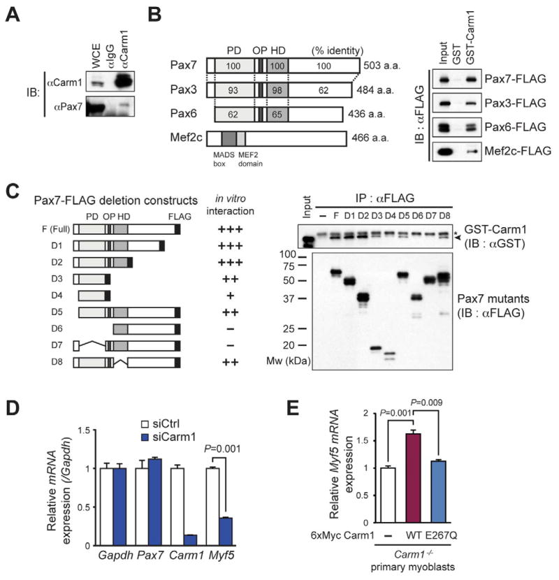
We identified Carm1/Prmt4 as a candidate Pax7 interacting protein following isolation of the Pax7 protein complex from primary myoblasts by tandem affinity purification and tandem mass spectrometry. The interaction between endogenous Pax7 and Carm1 was confirmed by co-immunoprecipitation analysis using Carm1 antibody in primary myoblasts (Figure 1A).
Figure 1. Carm1 interacts with Pax7 and regulates Myf5 expression, see also Figure S1.
(A) Co-immunoprecipitation of endogenous Pax7 with Carm1 from primary myoblasts. Endogenous Pax7 is detectable by immunoblotting when Carm1-associated proteins are immunoprecipitated with anti-Carm1 antibody from whole cell extract (WCE).
(B) Interactions between Carm1 and indicated FLAG tagged proteins were detected by in vitro GST pull-down assays. Schematic representation of mouse Pax7, Pax3, Pax6 and Mef2c were shown in left panel. Percentage identity between the paired domain (PD) or homeo domain (HD) of these proteins are indicated.
(C) Mapping of the Carm1-interacting domain of Pax7 to the N-terminal region containing the paired domain by in vitro binding (right). The left panel shows the Pax7 deletion series. PD; paired domain, OP; octa-peptide motif, HD; homeo box domain; Asterisk, nonspecific band.
(D) Depletion of Carm1 with siRNA results in decreased expression of Myf5. RT-qPCR analysis from control (siCtrl) and Carm1 (siCarm1) siRNA transfected primary myoblasts (n=3, error bars represent s.e.m.).
(E) Catalytically active Carm1 rescues Myf5 expression levels in Carm1-deficient myoblasts. RT-qPCR (right) analysis from mock, 6xMyc-Carm1 WT and E267Q mutant over-expressing Carm1−/− primary myoblasts. (n=3, error bars represent s.e.m.)
To determine whether Carm1 interacts directly with Pax7, purified recombinant, glutathione S-transferase (GST) tagged Carm1 and 3xFLAG tagged Pax7 (Pax7-FLAG) expressed in Sf9 cells were used in a GST binding assay (Figures 1B, S1A and S1B). We observed that GST-Carm1, but not GST alone, associated with Pax7-FLAG, demonstrating a direct interaction between Pax7 and Carm1. We also found that Carm1 binds Pax3 and Pax6, and confirmed the known interaction between Carm1 and Mef2c.
To identify the region of Pax7 that binds Carm1, we performed an in vitro binding experiment between a deletion series of Pax7-FLAG proteins and GST-Carm1. Notably, the N-terminal region of Pax7, containing the paired domain, was sufficient to bind Carm1 (Figure 1C). Therefore, these data suggest that the interaction with Carm1 is conserved among paired box domain proteins.
To investigate whether Carm1 has a role in regulating Pax7 activity, we assessed the expression level of Myf5 following transfection of Carm1 siRNA into primary myoblasts and confirmed the knockdown by qRT-PCR and western blot (Figure 1D and S1C). Strikingly, depletion of Carm1 with siRNA resulted in a marked decrease in expression of Myf5 mRNA (Figure 1D). Carm1 knockout (Carm1−/−) primary myoblasts generated from tamoxifen treated Pax7-CreERT2:Carm1flox/flox mice also displayed reduced levels of Myf5 expression relative to cells infected with retrovirus expressing wild type (WT) Carm1 (Figure 1E and S1D, See EXPERIMENTAL PROCEDURE). By contrast, infection with retrovirus expressing a catalytic mutant (E267Q) of Carm1 did not restore Myf5 mRNA levels (Figure 1E) (Lee et al., 2002). Ectopic expression of Carm1 WT in Carm1−/− primary myoblasts restored arginine methylation as detected by anti-histone H3 asymmetric di-methyl R17 (αH3R17me2a) antibody, but not in mock or Carm1 E267Q mutant infected Carm1−/− primary myoblasts (Figure S1D) (Yadav et al., 2003).
Overexpression of Pax7 in primary myoblasts results in up-regulation of Myf5 mRNA level (McKinnell et al., 2008). However, we found that overexpression of Pax7 in Carm1-deficient primary myoblasts was unable to induce Myf5 up-regulation, while simultaneous ectopic expression of Carm1 WT rescued Myf5 induction (see below). Taken together, these results are consistent with the hypothesis that Carm1 regulates the ability of Pax7 to activate Myf5 transcription.
Carm1 specifically methylates Pax7 in amino terminus domain
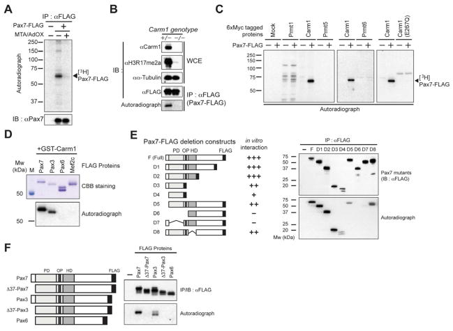
To evaluate the possibility of that Carm1 directly methylates Pax7, we conducted an in vivo methylation assay using the primary myoblasts ectopically expressing FLAG-tagged Pax7 (Pax7-FLAG) (Figure 2A). Ectopically expressed Pax7 was immunoprecipitated by anti-FLAG antibody and de novo methylation determined by autoradiography. At the same time, Prmt inhibitors, 5′-deoxy-5′-(methylthio) adenosine (MTA) and adenosine-2′, 3′-dialdehyde (Adox) were added into labeling media to inhibit arginine methylation. Since methylation of Pax7 was not evident in the presence of MTA/Adox, we conclude that Pax7 is methylated by an arginine methyltransferase in vivo.
Figure 2. Carm1 specifically methylates Pax7, see also Figure S2.
(A) Autoradiography and western blot analysis of immunoprecipitated Pax7-FLAG from in vivo [3H]-methionine labeled primary myoblasts cultured in the presence or absence of Carm1 inhibitor MTA/Adox. [3H]-labeled methylation of Pax7 was detected by autoradiography (top).
(B) Autoradiography and western blot analysis of immunoprecipitated Pax7-FLAG antibody from in vivo [3H]-methionine labeled Carm1-deficient primary myoblasts.
(C) Autoradiography of in vitro methylation assay of recombinant Pax7 with immunoprecipitated 6xMyc-Prmts.
(D) Autoradiography and protein staining of in vitro methylation assay for FLAG-tagged recombinant proteins with GST-Carm1.
(E) Domain mapping of the Pax7 region methylated by Carm1. C-terminal FLAG tagged Pax7 deletion constructs (left) were ectopically expressed in HEK293T cells and purified by anti-FLAG M2 agarose. Methylation of these constructs was analyzed using an in vitro methylation assay with purified GST-Carm1 and S-adenocyl [3H]-AdoMet and subsequently detected by autoradiography (right bottom). FLAG tagged Pax7 deletion mutants were detected by western blot analysis using anti-FLAG antibody (right top). Relative intensity methylation for Pax7 deletion mutants are indicated by + or − (+++ > ±).
(F) In vitro methylation assay for N-terminal 37 a.a. deleted Pax7, Pax3 and Pax6. C-terminal FLAG tagged Pax7, Pax3 and Pax6 constructs (left) were ectopically expressed in HEK293T cells and purified by anti-FLAG M2 agarose. Methylation of these proteins was analyzed by in vitro methylation assay using purified GST-Carm1 and detected by autoradiography (bottom right). FLAG tagged proteins were detected by western blot analysis using anti-FLAG antibody (top right).
To assess whether the in vivo methylation of Pax7 was ascribable to Carm1, we performed the same in vivo methylation assay using Cre-deleted Carm1 heterozygote (Carm1+/−) and Carm1−/− primary myoblasts overexpressing Pax7-FLAG. Importantly, our data demonstrated that Pax7 methylation is Carm1-dependent (Figure 2B, See EXPERIMENTAL PROCEDURE).
To extend this analysis, we performed in vitro methyltransferase assays for Pax7 using immunoprecipitated 6xMyc tagged Prmts including Carm1, Carm1 E267Q mutant, Prmt1, Prmt5, and Prmt6. The methyltransferase activity of the purified Prmts was confirmed by histone methyltransferase assays (Figure S2). Notably, this experiment demonstrated that only Carm1 is capable of methylating Pax7 (Figure 2C).
To understand the Carm1 substrate specificity, we applied an in vitro methylation assay using Carm1 binding proteins, including Pax7, Pax3, Pax6, and Mef2c shown in Figure 1B as substrates (Figure 2D). Interestingly, unlike the in vitro binding assay, specific methylation was detected using Pax7 and Pax3 as substrates, but not Pax6 and Mef2c. Therefore, Pax7 and Pax3 are specific substrates of Carm1, and interacting proteins are not necessarily always a substrate.
Although other Prmts are known to methylate arginine residues within glycine-argininerich motifs (GAR), the consensus sequence for Carm1 methylation has not been fully defined (Bedford and Clarke, 2009). To identify the arginines in Pax7 that are methylated by Carm1, we first performed in vitro methylation assays using the deletion series of Pax7 (Figure 2E). Significant methylation was detected in the N-terminal 1–37 a.a. of Pax7, with weaker methylation in the paired domain. Methylation was also detected in N-terminal 1–37 a.a. deleted Pax3, suggesting the N-terminal 37 a.a. of both Pax7 and Pax3, contain arginine residues that are methylated by Carm1 (Figure 2F).
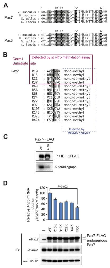
Examination of the N-terminal region among mouse paired-box transcription factors revealed a highly conserved region in Pax7 and Pax3 containing four arginine residues at position 10, 13, 22, and 37 (Figure S3A). The region also contained a proline-glycine-methionine (PGM) motif, which has been noted in Carm1 substrates along with the arginine residues (Figure S3B) (Bauer et al., 2002; Lee et al., 2002; Bedford et al., 2009; Sims et al., 2011). These arginine residues and PGM motifs are well conserved across vertebrate Pax7 and Pax3 proteins (Figure 3A). Liquid chromatography-tandem mass spectrometry analysis of immunoprecipitated Pax7-FLAG protein from Pax7-FLAG overexpressing primary myoblasts confirmed Arg10, 13, 22 and 37 as sites of methylation within the N-terminal domain and identified an additional 10 arginines that are also methylated (Figures 3B, S3C–D and Table S1).
Figure 3. Methylation of Pax7 by Carm1 within the N-terminal PGM motif is required for Myf5 induction, see also Figure S3.
(A) Amino acid sequence alignment of N-terminal 37 a.a. in Pax7 and Pax3 among the indicated species. Conserved arginine residues in Pax7 and Pax3 are highlighted in black and proline glycine methionine rich (PGM) motifs are highlighted in gray.
(B) Methylated arginines in Pax7 were identified by in vitro methylation assay or LC-MS/MS (lower). The PGM motif is highlighted in gray (proline, glycine and methionine) and arginine is highlighted in black.
(C) Autoradiography and western blot of immunoprecipitated Pax7-FLAG WT and 4RK mutant from in vivo [3H]-methionine labeled primary myoblasts.
(D) RT-qPCR and Western blot of primary myoblasts over-expressing Pax7-FLAG series of arginine mutants. Relative Myf5 mRNA induction is normalized to the amount of Pax7 mRNA in each cell line (n=3, error bars represent s.e.m.).
To assess the biological significance of this methylation, we substituted arginine residues R10, R13, R22 and R37 with lysine by site directed mutagenesis and performed in vitro methylation assays. When all four arginines were mutated in Pax7, we found a marked decrease in the level of methylation (4RK mutant: (R10K/R13K/R22K/R37K) (Figure S3E). Similar result was observed following in vitro methylation using Pax7-Pax6 chimeric protein containing 1–37aa from Pax7 4RK fused with Pax6 (Figure S3G). Importantly, in vivo methylation was markedly decreased in Pax7 4RK mutant when overexpressed in primary myoblasts (Figure 3C and S3F). Finally, overexpression of the Pax7 4RK mutant was unable to induce Myf5 transcription in the same manner as Pax7 WT (Figure 3D). Therefore, we conclude that R10, R13, R22, and R37 represent the major sites of methylation in Pax7 and are required for Pax7 function in vitro and in vivo.
Carm1 methylation of Pax7 does not affect DNA binding activity
To assess whether Carm1 directed methylation of Pax7 effects DNA binding, we conducted performed electrophoretic mobility shift assays (EMSA). WT and 4RK mutant of Pax7-FLAG proteins were purified from E. coli, and thus are not methylated on arginine residues. We then performed EMSA experiments using in vitro methylated or unmethylated Pax7 WT or 4RK mutant on Pax3/7 binding element from the −57.5 Kb Myf5 regulatory element (Figures S4A–C) (Bajard et al., 2006; McKinnell et al., 2008). Remarkably, EMSA experiments indicated that the DNA binding activity of Pax7 WT and 4RK mutant was unaffected by methylation (Figures S4D–E).
In addition, a one-hybrid experiment was performed by co-transfection of a C-terminal VP16 fused Pax7 WT or 4RK mutant with a luciferase reporter plasmid carrying the same DNA sequence as the EMSA experiment in C3H10T1/2 cells. These experiments indicated that the 4RK mutant exhibited unaltered activity (Figure S4F). These data demonstrate that DNA-binding by Pax7 is unaffected by Carm1 methylation and suggest the possibility that Pax7 cofactor recruitment is affected.
Carm1 methylation of Pax7 regulates binding of MLL1/2
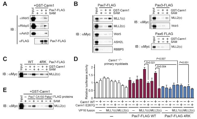
Pax7 recruits the ASH2L:WDR5:MLL1/2:RBBP5 histone methyltransferase (HMT) complex to regulatory elements on target genes such as Myf5 to activate transcription (McKinnell et al., 2008). Therefore, we investigated whether arginine methylation of Pax7 by Carm1 regulates recruitment of the HMT complex. The binding of methylated Pax7 to individual complex components was assessed by in vitro pull-down assays using methylated or unmethylated Pax7, with nuclear extracts, and the interactions assessed by western blot analysis using specific antibodies against the components of the HMT complex. Strikingly, we observed that methylated Pax7, but not unmethylated Pax7, bound the HMT complex (Figure 4A).
Figure 4. Direct interaction between arginine methylated Pax7 and MLL1/2, see also Figure S4.
(A) Methylated Pax7 interacts with the MLL complex. In vitro binding assays were performed using in vitro methylated Pax7 and nuclear extract from primary myoblasts.
(B) In vitro binding assay using in vitro methylated Pax7, Pax3 or Pax6 and in vitro translated 6xMyc-MLL1(c), MLL2(c), Wdr5, ASH2L or RBBP5.
(C) N-terminal Pax7 arginine methylation is required for interaction with MLL2. In vitro binding assays were performed using methylated Pax7 and in vitro translated 6xMyc-MLL2(c). Interactions were detected by western blot analysis (a–c).
(D) Pax7 and MLL1 or MLL2 interactions require Carm1. A three hybrid assay was performed by transfecting plasmid expressing Pax7, and/or VP16-MLL1(c), or MLL2(c), and/or 6xMyc-Carm1, with luciferase reporter plasmid containing Myf5 −57.5 kb promoter and renilla internal control reporter plasmid DNA into Carm1-deficient primary myoblasts. The interactions were detected as the relative luciferase activity compared to mock transfected control (n=3, error bars represent s.e.m.).
(E) In vitro binding assay between proteins with Carm1 arginine methylated PGM motifs, and in vitro translated 6xMyc-MLL2(c).
To identify which of the complex components was directly interacting with methylated Pax7, we performed the in vitro binding assay using methylated Pax7 and in vitro translated complex core components: C-terminal cleavage forms of MLL1/2; MLL1(c) and MLL2/TRX2(c), and ASH2L, RBBP5, and Wdr5 (Capotosti et al., 2007). We observed methylation dependent interaction only with the catalytic core component, MLL1(c) and MLL2(c), but not with any other components. Similarly, this interaction was noted with methylated Pax3, but not with Pax6 (Figure 4B). Moreover, methylation at R10, R13, R22 and R37 of Pax7 was required for this interaction, as the Pax7 4RK mutant did not bind MLL1/2 in the in vitro binding assay (Figures 4C and S3F).
To establish that the interaction between MLL1/2 and Pax7 is regulated by Carm1 in vivo, we performed a three-hybrid assay where Pax7-FLAG (WT or 4RK), VP16-MLL1(c) or VP16-MLL2(c), and 6Myc-Carm1 (WT or E267Q mutant) expression vectors using the Myf5 −57.5 kb luciferase reporter, were co-transfected into Cre-deleted Carm1−/− primary myoblasts (Figure 4D). Our results demonstrate that Carm1 methylates Pax7 to regulate the recruitment of the MLL complex to Pax7 bound regulatory sequences such as Myf5 gene through direct binding to the C-terminal forms of MLL1 and MLL2.
Several studies have recently demonstrated that Tudor domains can specifically recognize arginine methylated non-histone proteins. For example, the binding of the SMN Tudor domain to arginine methylated CA150, and TDRD3 to arginine methylated RNApolII CTD (Cheng et al., 2007; Cote et al., 2005; Sims et al., 2011). Moreover, TDRD3 contains a reader domain that specifically recognizes H3R17me2a and H4R3me2a marks that have been deposited by Carm1 or PRMT1 (Yang et al., 2010). Notably, the C-terminal forms of MLL1/2 do not contain a Tudor domain. Therefore, we assessed whether the C-terminal form of MLL2 recognizes other proteins that are methylated on arginine by Carm1, CA150 and Pabp1 (Chen et al., 2002; Lee et al., 2002). We performed an in vitro binding assay with arginine methylated or unmethylated Pax7-FLAG, CA150-FLAG or Pabp1-FLAG, and in vitro synthesized MLL2(c). Notably, we found that the C-terminal form of MLL2 specifically bound to methylated Pabp1, but not to CA150 (Figures 4E, S4G–H). Taken together, these data indicate that the C-terminal form of MLL2 contains a novel-binding module that specifically mediates interactions with a subset of proteins that are methylated on arginine residues.
Methylation of Pax7 is required for H3K4me3 enrichment and recruitment of MLL by Pax7 to the Myf5 promoter
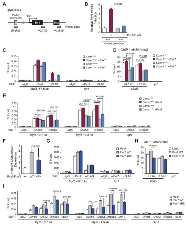
Our experiments establish that Carm1 methylates specific arginines in the amino-terminal region of Pax7. Once methylated, Pax7 is then specifically bound by MLL1/2 and the HMT complex assembled. To characterize the assembly process on chromatin, we examined Carm1 dependent MLL complex recruitment and the H3K4me3 enrichment on Myf5 +0.7 kb and +1.5 kb regions from TSS (transcription start site) and Pax7 binding on enhancer region (−57.5 kb) binding by ChIP-qPCR analysis (Figure 5A) (McKinnell et al., 2008).
Figure 5. Carm1 methylation of Pax7 is required for H3K4me3 enrichment and recruitment of the MLL complex to the Myf5 proximal promoter, see also Figure S5.
(A) Location of primer sets for ChIP-qPCR analysis of Myf5 promoter. TSS; transcription start site.
(B) RT-qPCR analysis of Myf5 mRNA expression.
(C) DNA binding activity of Pax7 on −57.5 kb Myf5 promoter in mock or Pax7-FLAG overexpressing Carm1-deleted primary myoblasts was determined by ChIP-qPCR analysis using anti-Pax7 anti-body or FLAG M2 antibody.
(D) ChIP-qPCR analysis of H3K4me3 enrichment.
(E) ChIP-qPCR analysis of MLL complex recruitment.
(n=3, error bars represent s.e.m., P-values are indicated in individual analysis)
(F) RT-qPCR analysis of Myf5 mRNA expression in Pax7 WT or 4RK mutant over-expressing primary myoblasts.
(G) DNA binding activity of Pax7 on −57.5 kb Myf5 promoter in mock, Pax7-FLAG WT or 4RK mutant over-expressing primary myoblasts was determined by ChIP-qPCR analysis using anti-Pax7 antibody or -FLAG M2 anti-body.
(H) ChIP-qPCR analysis of H3K4me3 enrichment in Pax7 WT or 4RK mutant over-expressing primary myoblasts.
(I) ChIP-qPCR analysis of MLL complex recruitment, in Pax7 WT or 4RK mutant overexpressing primary myoblasts.
(n=3, error bars represent s.e.m., P-values are indicated in individual analysis)
Mock or Pax7 overexpressing Carm1+/− and Carm1−/− primary myoblasts were established by retrovirus infection (Figure S5A) and Myf5 mRNA expression detected by qRT-PCR (Figure 5B). As expected, Pax7 overexpression in Carm1+/− cells revealed a 5-fold induction of Myf5 mRNA, whereas this induction is reduced in Carm1−/− cells. Consistent with our observations that Carm1 does not regulate Pax7 DNA binding, we found similar Pax7 binding in the enhancer region in both cell populations (Figure 5C). Examining the proximal promoter of Myf5, the increased enrichment of Ash2l and Rbbp5 and H3K4me3 were consistently correlated with Pax7 overexpression in Carm1+/− primary myoblasts. By contrast, reduced binding of Ash2l and Rbbp5, and reduced enrichment of H3K4me3 were consistently observed in Carm1−/− primary myoblasts, in spite of Pax7 overexpression (Figure 5D and 5E).
We next investigated whether arginine methylation of Pax7 was directly required to recruit MLL complex components and remodel chromatin over the Myf5 gene. Mock, Pax7 WT and 4RK mutant overexpressing primary myoblasts were established by retrovirus infection and analyzed for Carm1 and Pax7 protein and Myf5 mRNA expression. Pax7 WT and 4RK mutant overexpressing primary myoblasts showed a similar extent of Pax7 and Carm1 protein expression (Figure S5B). However, Pax7 4RK mutant overexpression resulted in reduced Myf5 induction (Figure 5F). Even though Pax7 binding at the Myf5 −57.5kb element was not affected with the loss of the arginine methylation sites (Figure 5G), the level of MLL complex recruitment by the 4RK mutant and accumulation of H3K4me3 on Myf5 was similar to mock infected primary myoblasts (Figure 5H–I). These data support the idea that arginine methylation of Pax7 by Carm1 is required for MLL complex recruitment and subsequent H3K4me3 over the Myf5 promoter.
Carm1 is required for satellite cell function in vivo
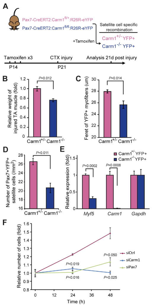
To examine the functional role of Carm1 in Pax7-expressing satellite cells, we used Pax7-CreERT2:Carm1fl/+:R26R-eYFP and Pax7-CreERT2:Carm1fl/fl:R26R-eYFP mice to conditionally inactivate Carm1. Moreover, simultaneous recombination at the R26R-eYFP locus allows us to analyze the functional capacities of Carm1+/− and Carm1−/− satellite cells by labeling their progeny with eYFP. Tamoxifen was injected to activate Pax7-CreERT2, which is expressed from the Pax7 promoter, leading to a tissue specific deletion of Carm1 at P14 to generate mice with Carm1+/−YFP+ and Carm1−/−YFP+ satellite cells. One week later, animals were injured with Cardiotoxin directly injected into the tibialis anterior (TA) muscle and analyzed after 21days (Figure 6A). Carm1−/− mice exhibited a marked regeneration deficit whereby injured muscles weighed ~25% less than the muscles from their heterozygous littermates (Figure 6B). Upon closer examination, YFP+ myofibers deriving from Carm1−/− YFP+ satellite cells had a significantly smaller minimum ferret diameter compared to those derived from Carm1+/− YFP+ satellite cells (Figure 6C and S6). Moreover, the number of Pax7+YFP+ satellite cells was also significantly reduced in Carm1−/− mice compared to those in the Carm1+/− mice (Figure 6D). To determine whether Carm1 regulate Myf5 expression in satellite cells, we used fluorescence activated cell sorting (FACS) to prospectively isolate Carm1+/−YFP+ and Carm1−/−YFP+ satellite cells from mice that were Tamoxifen treated at P14. Strikingly, we found that, in vivo, Myf5 expression is reduced by ~65% in Carm1−/− satellite cells (Figure 6E). This further demonstrates the requirement for Carm1 in satellite cells during regeneration.
Figure 6. In vivo function of Carm1 in satellite cells, see also Figure S6.
(A) Schematic of the conditional knockout model used (upper) and the timeline used for the regeneration studies (lower).
(B) Relative weight of injured TA muscles 21 days-post Cardiotoxin injury (n=3 per sample group; error bars represent s.e.m.).
(C) Minimum feret diameter of YFP+ fibers measured from immunostained TA muscle cross-sections (n>50 fibers per mouse; 3 biological replicates; error bars represent s.e.m.).
(D) Number of Pax7+YFP+ satellite cells observed in immunostained TA muscle cross-sections (n=3; error bars represent s.e.m.).
(E) qRT-PCR analysis of freshly sorted Carm1+/−YFP+ and Carm1−/−YFP+ satellite cells normalized to Gapdh expression (n=3; error bars represent s.e.m.)
(F) Growth curve of P21 juvenile primary myoblasts transfected with siCrtl, siCarm1 or siPax7 normalized to 0h (n=3; error bars represent s.e.m.)
Several studies have previously implicated Carm1 in terminal myogenic differentiation of myoblasts (Chen et al., 2002; Gao et al., 2010; Rudnicki lab, data not shown). Thus the observed regeneration deficit could be the result of an inability for Carm1−/−YFP+ satellite cells to undergo differentiation. Thus, we examined the role of Carm1 in proliferation conditions. Using low-passage primary myoblasts generated from FACS purified P21 juvenile Pax7-ZsGreen satellite cells, we perturbed either Carm1 or Pax7 expression by siRNA and monitored their rate of proliferation. Similar to siPax7, P21 myoblasts transfected with siCarm1 showed severe growth deficits (Figure 6F). This suggests that the reduction in Pax7+YFP+ satellite cells in Carm1−/− muscles after injury is due to a proliferation deficit.
Carm1 is required to activate Myf5 during asymmetric stem cell divisions
Arginine methylation of Pax7 by Carm1 is required for Myf5 induction. Therefore, we hypothesized that the de novo activation of Myf5 transcription that occurs following asymmetric satellite stem cell division requires MLL complex recruitment and that this induction is regulated by Carm1-mediated arginine methylation of Pax7. Satellite stem cells and their daughter satellite myogenic cells are readily discerned using Myf5-Cre and R26R-YFP alleles (Kuang et al., 2007). Satellite stem cells have never expressed Myf5-Cre and hence do not express R26R-YFP. By contrast, satellite myogenic cells have expressed Myf5-Cre and thus express R26R-YFP. Satellite stem cells give rise to satellite myogenic cells through asymmetric apical-basal cell divisions that are visualized during culture of isolated myofibers (Kuang et al., 2007).
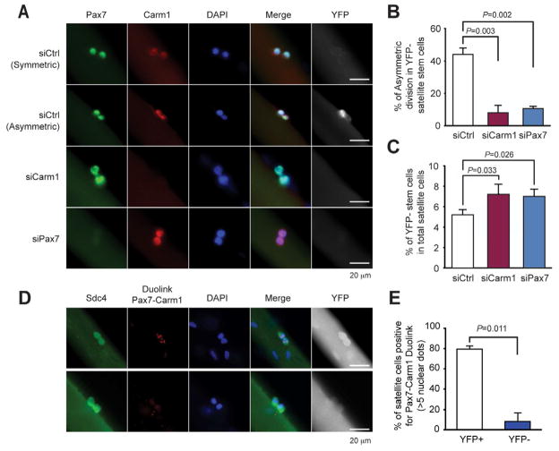
To analyze the requirement for Carm1 during asymmetric satellite stem cell divisions, Carm1 was depleted by siRNA knockdown in satellite cells on single myofibers from Myf5 Cre/R26R-YFP mice. As a control, Pax7 was knocked down in parallel experiments, to ascertain the requirement for Pax7 for the induction of Myf5 following asymmetric stem cell division. Strikingly, we observed that the percentage of asymmetric satellite stem cell divisions after 42h of culture was markedly reduced following siRNA transfection of siCarm1 or siPax7 (Figure 7A). We observed that 44% of stem cell divisions were asymmetric following transfection of scrambled siRNA (Figure 7B). By contrast, transfection of siCarm1 significantly reduced the proportion of asymmetric divisions to 8% (p=0.003). Similarly, transfection of siPax7 reduced the proportion of asymmetric cell divisions to 10% (p=0.002). Importantly, the total number of satellite cells was not changed (Figure S7A) but the overall proportion of satellite stem cells increased by about 30% (Figure 7C). Moreover, a similar reduction of asymmetric division in Pax7 knockdown satellite stem cells was detected by immunostaining using anti-CD34, -Pax7 and -YFP antibodies (data not shown).
Figure 7. Carm1 regulates Myf5 induction following satellite stem cell asymmetric cell division, see also Figure S7.
(A) Immunocytochemistry of Pax7, Carm1 and YFP in Ctrl, Carm1 or Pax7 siRNA treated muscle fibers isolated from Myf5-Cre/RosaYFP mice and cultured for 42h. Satellite stem cells are YFP− and satellite myogenic cells are YFP+ Pax7 (green); Carm1 (red), DAPI (blue); YFP (white); Scale bars represent 20μm.
(B) Increased numbers of satellite stem cells after Carm1 or Pax7 siRNA knock down.
(C) Asymmetric satellite stem cells divisions are markedly decreased following knock down of Carm1 or Pax7.
(D) Proximity ligation assay against Pax7 and Carm1 (red) counter stained for Sdc4 (green) to demarcate satellite cells, DAPI (blue) and YFP to distinguish satellite stem cells and myogenic satellite cells (white). Scale bars represent 20μm.
(E) Quantification of the proximity ligation assay. Duolink+ cells were identified as >5 punctate dots in the nucleus (as shown in panel D; upper) (n=4; error bars represent s.e.m.).
Pax7 and Carm1 are both expressed in YFP+ satellite myogenic cells and YFP− satellite stem cells. However, only YFP+ stem cells have an active Myf5 promoter. Using a proximity ligation assay (Duolink), we set out to examine whether Pax7-Carm1 interactions take place equally in both cell types. Importantly, we find that Pax7 and Carm1 only interact in the nucleus of YFP+ satellite myogenic cells (Figure 7E–F). By knocking down Carm1 with siRNA, we lose this interaction completely (Figure S7B). This correlation between Pax7-Carm1 interactions and Myf5 expression in satellite cells strongly supports the hypothesis that Carm1 methylation of Pax7 is required for Myf5 expression, and also suggests Carm1 is itself subject to regulation during asymmetric cell division.
Taken together, these data demonstrate a critical role for Carm1 in regulating Pax7 function in muscle stem cells. The presence of similar levels of Carm1 protein in the nuclei of satellite stem cells and satellite myogenic cells (Figure 7A), suggests that Carm1 activity is subject to post-translational regulation. Therefore, we conclude that the de novo activation of Myf5 transcription in committed satellite myogenic cells, following an asymmetric stem cell division, requires Carm1-mediated methylation of Pax7.
DISCUSSION
The molecular mechanism through which activation of genes that enforce lineage commitment following asymmetric stem cell divisions has long remained an open question. Our experiments identify Carm1 as the key factor that activates Pax7 during the commitment of satellite stem cells. We demonstrate that arginine methylation of Pax7 is required for MLL1/2 binding and HMT complex assembly. Moreover, we found that Carm1 binding to Pax7 is inhibited in satellite stem cells in order to maintain the stem cell state. Thus, Carm1 methylation of Pax7 functions as the molecular switch for the epigenetic activation of the myogenic determination gene Myf5.
Carm1 is a protein arginine methyltransferase that methylates histone R17 and 26 and has an activator function that is coupled with the SWI/SNF chromatin-remodeling complex (Xu et al., 2004). Histone H3 arginine methylation at R17 and 26 is enriched on transcriptional active genes and Carm1 regulated genes are enriched by H3R17me2 and H3R26me2 modifications in their promoter regions (Ma et al., 2001). We examined Carm1 localization and H3K17me2 enrichment on the −57.5 kb enhancer and +0.7 kb proximal promoter of Myf5 using ChIP-qPCR, and detected no significant enrichment in Carm1 binding nor H3K17me2 (data not shown). Therefore, Carm1 methylation of Pax7 versus that of histones H3, appears mechanistically independent of one another.
Carm1 has recently been demonstrated to regulate the establishment of cell polarity by changing the apical localization of the partition defective (Par)-complex to promote asymmetric cell division in early mouse embryos (Torres-Padilla et al., 2007; Parfitt et al., 2010). The asymmetric distribution of the Par-complex has been noted in asymmetric stem cell divisions in Drosophila, the vertebrate zygote and in adult stem cells (Suzuki et al., 2006). The Par-complex, comprised of Par-3/Par-6/atypical protein kinase C (aPKC), and participates in Numb phosphorylation and the polarized localization of phosphorylated Numb (Tajbakhsh et al., 2009). Our experiments indicate that activation of Myf5 transcription in the apical satellite cell requires Carm1 and Pax7. However, whether this process is mechanistically downstream of the Par complex or functions in a parallel manner remains to be addressed.
Equivalent nuclear expression of Carm1 is observed in both satellite stem cells and satellite myogenic cells, but can only bind to Pax7 in satellite myogenic cells. Therefore, it is reasonable to speculate that Carm1 is regulated by post-translational modification during satellite stem cell asymmetric cell division. Several groups have reported that Carm1 is regulated by phosphorylation during mitosis (Higashimoto et al., 2007; Feng et al., 2009). Phosphorylation of Carm1 at Ser217 inhibits arginine methylation activity by either diminished methyl donor binding or by promoting cytoplasmic localization. These experiments raise the possibility that Carm1 is phosphorylated in satellite stem cells thus inhibiting its activity. Alternatively, the interaction between Pax7 and Carm1 may be regulated during satellite stem cell asymmetric cell division.
The regeneration deficit observed when Carm1 is inactivated prior to injury-induced regeneration clear demonstrates the in vivo requirement of Carm1 in myogenesis. Although it has been previously reported that Carm1 interacts with Mef2C and other effectors to regulate terminal differentiation (Chen et al., 2002; Gao et al., 2010), we observe that quiescent satellite cells depleted of Carm1 exhibit markedly reduced Myf5 expression and myoblasts maintained in growth conditions proliferate poorly when they are transfected with siRNA against Carm1. In addition, reduced numbers of Pax7+YFP+ satellite cells were observed in Carm1−/− mice after injury. Therefore, we conclude that Carm1 is required for satellite stem cell activation of Myf5 transcription and hence for myoblast expansion.
Myf5 induction demarcates the entry of satellite stem cells into the myogenic program. In embryonic myogenesis, Pax3, Wnt and Shh signaling pathways have been implicated in regulating Myf5 transcription (Bajard et al., 2006; Tajbakhsh et al., 1998; Borycki et al., 1999). However, the signaling pathways regulating Myf5 expression in adult myogenesis remains undefined. Previously, we reported that Pax7 directly binds to Myf5 −57.5 kb promoter and recruits the MLL1/2 complex to epigenetically regulate Myf5 transcription in adult satellite cells (McKinnell et al., 2008). We have also identified a role for non-canonical Wnt7a/Fzd7 signaling through the planar-cell-polarity pathway to drive symmetric stem cell expansion (Le Grand et al., 2009). Whether Wnt7a/Fzd7 signaling has a direct or indirect role in regulating Carm1 activity remains to be determined.
Histone methylation is an important mechanism in the epigenetic regulation of stem cell fate decisions, and the recruitment of HMTs is highly regulated (Ruthenburg et al., 2007; Lim et al., 2009). Our study has established a new level of regulation for the recruitment of HMTs on proximal promoter of transcriptionally active genes. Our experiments demonstrate that Carm1 methylation of arginines in the amino terminus of Pax7 creates an interface that is specifically recognized and bound by the C-terminal cleavage forms of MLL1/2 and subsequently the MLL HMT complex. This interaction recruits the MLL HMT complex to the proximal promoter sequences of Myf5, leading to its transcriptional induction.
Recent findings implicate the interaction between histones and effector proteins through methylation as a crucial step in the translation of the histone code in influencing downstream biological processes (Kouzarides et al., 2007; Yun et al., 2011). PHD and Tudor royal domain comprising chromo, PWWP and MBT domain recognize histone lysine methylation at H3K4, K9, K27, K36 and H4K20 (Chen et al., 2011). H3R17me2a and H4R3me2a marks are recognized by a reader domain in TDRD3 (Yang et al., 2010). Notably, PHD finger in nucleosome remodeling factor (NURF), an ISWI-containing ATP-dependent chromatin-remodeling complex, recognizes H3K4me3 and this recognition is coupled with NURF-mediated ATP-dependent chromatin remodeling to maintain Hox gene expression patterns during development (Wysocka et al., 2006).
Protein-protein binding of non-histone proteins is also mediated by binding of particular domains to methylated sequences (Bedford and Clarke, 2009; Sims et al., 2008). For example, lysine methylated p53 and arginine methylated CA150 and RNA polII are recognized by the Tudor domains in 53BP1, SMN, and TDRD3, respectively to specify interactions and mediate downstream events (Huang et al., 2007; Cheng et al., 2007; Sims et al., 2011). Notably, our discovery that the recognition and binding to arginine methylated Pax7 and Pabp1 by the C-terminal region of MLL1/2 reveals the existence of a new class of reader module for the specific recognition of arginine methylated proteins.
Our experiments have defined the C-terminal region of MLL1/2 as containing a novel module for the specific recognition of arginine methylation on Pax7 to effect sequence-specific protein-protein binding. Moreover, our findings demonstrate that Carm1 directed methylation of Pax7 functions as the molecular switch for the epigenetic activation of the myogenic determination gene Myf5 following the asymmetric division of a satellite stem cell. Together, these experiments provide important insight into the molecular mechanisms that regulate the specification of stem cells and the regulation of gene expression during asymmetric stem cell divisions..
EXPERIMENTAL PROCEDURES
Detail experimental procedures are described in SUPPLEMENTAL EXPERIMENTAL PROCEDURES
Cell culture
Primary myoblasts were cultured as previously described5. For protein expression or siRNA knockdown in primary myoblasts, cells were infected by retrovirus with polybrene or transfected by LipofectAmine RNAiMAX reagent (Invitrogen).
Recombinant protein expression and purification
C-terminally 3xFLAG-tagged Pax7, Pax3, Pax6, CA150, Pabp1, GST-Carm1 and 6xMyc tagged Prmts were expressed in Sf9 cells, BL21-Codon-Plus(DE3)-RIL cells or HEK293T cells and purified by affinity purification.
In vitro binding assay
GST fusion protein or FLAG tagged fusion protein was mixed with either the indicated proteins or nuclear extract in binding buffer.
In vitro methyltransferase assay
The purified methyltransferases were mixed with core histones (Upstate) or purified substrate proteins in the presence of S-adenylyl [3H]-AdoMet. [3H]-labeled proteins were resolved by SDS-PAGE and detected by autoradiography.
In vivo labeling of primary myoblasts
Mock or Pax7-FLAG over-expressing primary myoblasts were incubated in the presence of L-[methyl-3H] Methionine. Immunoprecipitated Pax7 was analyzed by western blot analysis and autoradiography.
Luciferase reporter assay
The indicated plasmids were transfected into primary myoblasts or C3H10T1/2 cells with the firefly and renilla luciferase reporters. Reporter activation was measured by LUMIstar OPTIMA (BMG Labtech) using Dual-Luciferase Reporter Assay System (Promega).
Chromatin immunoprecipitation (ChIP)
The cultured primary myoblasts were cross-linked with either 1 % formaldehyde or 5 mM Dimethyl 3,3′-dithiobispropionimidate 2HCl (Thermo). Cross-linked cells were sheared by Bioruptor (Diagenode) and immunoprecipitated with the indicated antibodies. Immunoprecipitated DNA was de-cross-linked, recovered by phenol/chloroform extraction and analyzed by quantitative PCR using CFR384 (Bio-rad).
Mice and myofiber experiment
Myf5-Cre/ROSA26-YFP mouse was previously described (Kuang et al., 2007). Single muscle fibers were isolated from the EDL muscle of 6–8 week old Myf5-Cre/RosaYFP mice (SV129) and transfected with siRNA as described previously (Kuang et al., 2007; Le Grand et al., 2009). Pax7-CreERT2 homozygote mouse and Carm1flox/flox mouse were crossed to obtain Pax7-CreERT2:Carm1+/flox heterozygous (Carm1+/−) and Pax7-CreERT2:Carm1flox/flox homozygous (Carm1−/−) (Yadav et al., 2003; Nishijo et al., 2009). Tamoxifen treatment and injury experiments were conducted as described previously following the University of Ottawa regulations for animal care and handling (Lepper et al., 2009).
Immunofluorescence and proximity ligation assay
Immunohistochemistry were conducted as described previously (Kuang et al., 2007; LeGrand et al., 2009). Duolink (OLink Bioscience) was used according to manufacturer’s recommendations to visualize the in vivo interaction between Pax7 and Carm1.
Supplementary Material
HIGHLIGHT.
Carm1 methylates Pax7 at specific arginines within the N-terminus.
Methylated Pax7 directly binds to C-terminal cleavage forms of MLL1/2.
Methylated Pax7 recruits ASH2L:MLL1/2:WDR5:RBBP5 H3K4 methyltransferase complex.
Pax7-Carm1 is required for Myf5 expression following asymmetric stem cell division.
Acknowledgments
We thank Drs. F. Jeffrey Dilworth and Marjorie Brand for critical reading of the manuscript, Jennifer Ritchie for expert technical assistance on animal experiments, Dr. Lawrence Puente for mass spectrometry analysis, Paul Oleynick for fluorescent-activated cell sorting, David Wilson for histological analysis and Drs. Chandra Prakash Chaturvedi and Carmen Palii, and Rudnicki lab members for helpful discussions. We thank Dr. Hess L Jay for MLL1 cDNA, Dr. Jocelyn cote for SMN-Tdr and TDRD3-Tdr plasmids, and Dr. F Jeffrey Dilworth for FLAG-tagged Mef2c protein. We thank Dr. Mark Gillespie and Dr. Jeffrey A Ranish for preliminary identification of Pax7 methylation site by mass spectrometry analysis. Y.K. is supported by fellowships from Uehara Memorial Foundation Scholarship and Ontario Research Fund. Y.X.W is supported by a fellowship from Ontario Research Fund. M.A.R. holds the Canada Research Chair in Molecular Genetics and is an International Research Scholar of the Howard Hughes Medical Institute. M.A.R. is supported by grants from the National Institutes of Health (R01AR044031), the Howard Hughes Medical Institute, the Canadian Institutes of Health Research, the Muscular Dystrophy Association, the Stem Cell Network and the Canada Research Chair Program. M.B. is supported by NIH grant DK6224.
Footnotes
Publisher's Disclaimer: This is a PDF file of an unedited manuscript that has been accepted for publication. As a service to our customers we are providing this early version of the manuscript. The manuscript will undergo copyediting, typesetting, and review of the resulting proof before it is published in its final citable form. Please note that during the production process errorsmaybe discovered which could affect the content, and all legal disclaimers that apply to the journal pertain.
References
- Abou-Khalil R, Le Grand F, Pallafacchina G, Valable S, Authier FJ, Rudnicki MA, Gherardi RK, Germain S, Chretien F, Sotiropoulos A, et al. Autocrine and paracrine angiopoietin 1/Tie-2 signaling promotes muscle satellite cell self-renewal. Cell Stem Cell. 2009;5:298–309. doi: 10.1016/j.stem.2009.06.001. [DOI] [PMC free article] [PubMed] [Google Scholar]
- Bajard L, Relaix F, Lagha M, Rocancourt D, Daubas P, Buckingham ME. A novel genetic hierarchy functions during hypaxial myogenesis: Pax3 directly activates Myf5 in muscle progenitor cells in the limb. Genes Dev. 2006;20:2450–2464. doi: 10.1101/gad.382806. [DOI] [PMC free article] [PubMed] [Google Scholar]
- Bauer UM, Daujat S, Nielsen SJ, Nightingale K, Kouzarides T. Methylation at arginine 17 of histone H3 is linked to gene activation. EMBO Rep. 2002;3:39–44. doi: 10.1093/embo-reports/kvf013. [DOI] [PMC free article] [PubMed] [Google Scholar]
- Bedford MT, Clarke SG. Protein arginine methylation in mammals: who, what, and why. Mol Cell. 2009;33:1–13. doi: 10.1016/j.molcel.2008.12.013. [DOI] [PMC free article] [PubMed] [Google Scholar]
- Bertani S, Sauer S, Bolotin E, Sauer F. The noncoding RNA Mistral activates Hoxa6 and Hoxa7 expression and stem cell differentiation by recruiting MLL1 to chromatin. Mol Cell. 2011;43:1040–1046. doi: 10.1016/j.molcel.2011.08.019. [DOI] [PMC free article] [PubMed] [Google Scholar] [Retracted]
- Bishcoff R. In: The satellite cell and muscle regeneration. Myology AG, Engel, Franzini-Armstrong C, editors. New York: McGraw-Hill; 1994. pp. 97–118. [Google Scholar]
- Borycki AG, Brunk B, Tajbakhsh S, Buckingham M, Chiang C, Emerson CP., Jr Sonic hedgehog controls epaxial muscle determination through Myf5 activation. Development. 1999;126:4053–4063. doi: 10.1242/dev.126.18.4053. [DOI] [PubMed] [Google Scholar]
- Capotosti F, Hsieh JJ, Herr W. Species selectivity of mixed-lineage leukemia/trithorax and HCF proteolytic maturation pathways. Mol Cell Biol. 2007;27:7063–7072. doi: 10.1128/MCB.00769-07. [DOI] [PMC free article] [PubMed] [Google Scholar]
- Charge SB, Rudnicki MA. Cellular and molecular regulation of muscle regeneration. Physiol Rev. 2004;84:209–238. doi: 10.1152/physrev.00019.2003. [DOI] [PubMed] [Google Scholar]
- Chen C, Nott TJ, Jin J, Pawson T. Deciphering arginine methylation: Tudor tells the tale. Nat Rev Mol Cell Biol. 2011;12:629–642. doi: 10.1038/nrm3185. [DOI] [PubMed] [Google Scholar]
- Chen D, Ma H, Hong H, Koh SS, Huang SM, Schurter BT, Aswad DW, Stallcup MR. Regulation of transcription by a protein methyltransferase. Science. 1999;284:2174–2177. doi: 10.1126/science.284.5423.2174. [DOI] [PubMed] [Google Scholar]
- Chen SL, Loffler KA, Chen D, Stallcup MR, Muscat GE. The coactivatorassociated arginine methyltransferase is necessary for muscle differentiation: CARM1 coactivates myocyte enhancer factor-2. J Biol Chem. 2002;277:4324–4333. doi: 10.1074/jbc.M109835200. [DOI] [PubMed] [Google Scholar]
- Cheng D, Cote J, Shaaban S, Bedford MT. The arginine methyltransferase CARM1 regulates the coupling of transcription and mRNA processing. Mol Cell. 2007;25:71–83. doi: 10.1016/j.molcel.2006.11.019. [DOI] [PubMed] [Google Scholar]
- Collins CA, Olsen I, Zammit PS, Heslop L, Petrie A, Partridge TA, Morgan JE. Stem cell function, self-renewal, and behavioral heterogeneity of cells from the adult muscle satellite cell niche. Cell. 2005;122:289–301. doi: 10.1016/j.cell.2005.05.010. [DOI] [PubMed] [Google Scholar]
- Cote J, Richard S. Tudor domains bind symmetrical dimethylated arginines. J Biol Chem. 2005;280:28476–28483. doi: 10.1074/jbc.M414328200. [DOI] [PubMed] [Google Scholar]
- Dacwag CS, Bedford MT, Sif S, Imbalzano AN. Distinct protein arginine methyltransferases promote ATP-dependent chromatin remodeling function at different stages of skeletal muscle differentiation. Mol Cell Biol. 2009;29:1909–1921. doi: 10.1128/MCB.00742-08. [DOI] [PMC free article] [PubMed] [Google Scholar]
- Feng Q, He B, Jung SY, Song Y, Qin J, Tsai SY, Tsai MJ, O’Malley BW. Biochemical control of CARM1 enzymatic activity by phosphorylation. J Biol Chem. 2009;284:36167–36174. doi: 10.1074/jbc.M109.065524. [DOI] [PMC free article] [PubMed] [Google Scholar]
- Feng Q, Yi P, Wong J, O’Malley BW. Signaling within a coactivator complex: methylation of SRC-3/AIB1 is a molecular switch for complex disassembly. Mol Cell Biol. 2006;26:7846–7857. doi: 10.1128/MCB.00568-06. [DOI] [PMC free article] [PubMed] [Google Scholar]
- Gao X, Pan WS, Dai H, Zhang Y, Wu NH, Shen YF. CARM1 activates myogenin gene via PCAF in the early differentiation of TPA-induced rhabdomyosarcoma-derived cells. J Cell Biochem. 2010;110:162–170. doi: 10.1002/jcb.22522. [DOI] [PubMed] [Google Scholar]
- Guenther MG, Jenner RG, Chevalier B, Nakamura T, Croce CM, Canaani E, Young RA. Global and Hox-specific roles for the MLL1 methyltransferase. Proc Natl Acad Sci U S A. 2005;102:8603–8608. doi: 10.1073/pnas.0503072102. [DOI] [PMC free article] [PubMed] [Google Scholar]
- Higashimoto K, Kuhn P, Desai D, Cheng X, Xu W. Phosphorylation-mediated inactivation of coactivator-associated arginine methyltransferase 1. Proc Natl Acad Sci U S A. 2007;104:12318–12323. doi: 10.1073/pnas.0610792104. [DOI] [PMC free article] [PubMed] [Google Scholar]
- Huang J, Sengupta R, Espejo AB, Lee MG, Dorsey JA, Richter M, Opravil S, Shiekhattar R, Bedford MT, Jenuwein T, et al. p53 is regulated by the lysine demethylase LSD1. Nature. 2007;449:105–108. doi: 10.1038/nature06092. [DOI] [PubMed] [Google Scholar]
- Kassar-Duchossoy L, Gayraud-Morel B, Gomes D, Rocancourt D, Buckingham M, Shinin V, Tajbakhsh S. Mrf4 determines skeletal muscle identity in Myf5:Myod doublemutant mice. Nature. 2004;431:466–471. doi: 10.1038/nature02876. [DOI] [PubMed] [Google Scholar]
- Kassar-Duchossoy L, Giacone E, Gayraud-Morel B, Jory A, Gomes D, Tajbakhsh S. Pax3/Pax7 mark a novel population of primitive myogenic cells during development. Genes Dev. 2005;19:1426–1431. doi: 10.1101/gad.345505. [DOI] [PMC free article] [PubMed] [Google Scholar]
- Kouzarides T. Chromatin modifications and their function. Cell. 2007;128:693–705. doi: 10.1016/j.cell.2007.02.005. [DOI] [PubMed] [Google Scholar]
- Kowenz-Leutz E, Pless O, Dittmar G, Knoblich M, Leutz A. Crosstalk between C/EBPbeta phosphorylation, arginine methylation, and SWI/SNF/Mediator implies an indexing transcription factor code. EMBO J. 2010;29:1105–1115. doi: 10.1038/emboj.2010.3. [DOI] [PMC free article] [PubMed] [Google Scholar]
- Kuang S, Charge SB, Seale P, Huh M, Rudnicki MA. Distinct roles for Pax7 and Pax3 in adult regenerative myogenesis. J Cell Biol. 2006;172:103–113. doi: 10.1083/jcb.200508001. [DOI] [PMC free article] [PubMed] [Google Scholar]
- Kuang S, Kuroda K, Le Grand F, Rudnicki MA. Asymmetric self-renewal and commitment of satellite stem cells in muscle. Cell. 2007;129:999–1010. doi: 10.1016/j.cell.2007.03.044. [DOI] [PMC free article] [PubMed] [Google Scholar]
- Le Grand F, Jones AE, Seale V, Scime A, Rudnicki MA. Wnt7a activates the planar cell polarity pathway to drive the symmetric expansion of satellite stem cells. Cell Stem Cell. 2009;4:535–547. doi: 10.1016/j.stem.2009.03.013. [DOI] [PMC free article] [PubMed] [Google Scholar]
- Lee J, Bedford MT. PABP1 identified as an arginine methyltransferase substrate using high-density protein arrays. EMBO Rep. 2002;3:268–273. doi: 10.1093/embo-reports/kvf052. [DOI] [PMC free article] [PubMed] [Google Scholar]
- Lee YH, Koh SS, Zhang X, Cheng X, Stallcup MR. Synergy among nuclear receptor coactivators: selective requirement for protein methyltransferase and acetyltransferase activities. Mol Cell Biol. 2002;22:3621–3632. doi: 10.1128/MCB.22.11.3621-3632.2002. [DOI] [PMC free article] [PubMed] [Google Scholar]
- Lepper C, Conway SJ, Fan CM. Adult satellite cells and embryonic muscle progenitors have distinct genetic requirements. Nature. 2009;460:627–631. doi: 10.1038/nature08209. [DOI] [PMC free article] [PubMed] [Google Scholar]
- Lim DA, Huang YC, Swigut T, Mirick AL, Garcia-Verdugo JM, Wysocka J, Ernst P, Alvarez-Buylla A. Chromatin remodelling factor Mll1 is essential for neurogenesis from postnatal neural stem cells. Nature. 2009;458:529–533. doi: 10.1038/nature07726. [DOI] [PMC free article] [PubMed] [Google Scholar]
- Ma H, Baumann CT, Li H, Strahl BD, Rice R, Jelinek MA, Aswad DW, Allis CD, Hager GL, Stallcup MR. Hormone-dependent, CARM1-directed, argininespecific methylation of histone H3 on a steroid-regulated promoter. Curr Biol. 2001;11:1981–1985. doi: 10.1016/s0960-9822(01)00600-5. [DOI] [PubMed] [Google Scholar]
- McKinnell IW, Ishibashi J, Le Grand F, Punch VG, Addicks GC, Greenblatt JF, Dilworth FJ, Rudnicki MA. Pax7 activates myogenic genes by recruitment of a histone methyltransferase complex. Nat Cell Biol. 2008;10:77–84. doi: 10.1038/ncb1671. [DOI] [PMC free article] [PubMed] [Google Scholar]
- Montarras D, Morgan J, Collins C, Relaix F, Zaffran S, Cumano A, Partridge T, Buckingham M. Direct isolation of satellite cells for skeletal muscle regeneration. Science. 2005;309:2064–2067. doi: 10.1126/science.1114758. [DOI] [PubMed] [Google Scholar]
- Nishijo K, Hosoyama T, Bjornson CR, Schaffer BS, Prajapati SI, Bahadur AN, Hansen MS, Blandford MC, McCleish AT, Rubin BP, et al. Biomarker system for studying muscle, stem cells, and cancer in vivo. FASEB J. 2009;23:2681–2690. doi: 10.1096/fj.08-128116. [DOI] [PMC free article] [PubMed] [Google Scholar]
- Oustanina S, Hause G, Braun T. Pax7 directs postnatal renewal and propagation of myogenic satellite cells but not their specification. EMBO J. 2004;23:3430–3439. doi: 10.1038/sj.emboj.7600346. [DOI] [PMC free article] [PubMed] [Google Scholar]
- Parfitt DE, Zernicka-Goetz M. Epigenetic modification affecting expression of cell polarity and cell fate genes to regulate lineage specification in the early mouse embryo. Mol Biol Cell. 2010;21:2649–2660. doi: 10.1091/mbc.E10-01-0053. [DOI] [PMC free article] [PubMed] [Google Scholar]
- Relaix F, Montarras D, Zaffran S, Gayraud-Morel B, Rocancourt D, Tajbakhsh S, Mansouri A, Cumano A, Buckingham M. Pax3 and Pax7 have distinct and overlapping functions in adult muscle progenitor cells. J Cell Biol. 2006;172:91–102. doi: 10.1083/jcb.200508044. [DOI] [PMC free article] [PubMed] [Google Scholar]
- Rudnicki MA, Le Grand F, McKinnell I, Kuang S. The molecular regulation of muscle stem cell function. Cold Spring Harb Symp Quant Biol. 2008;73:323–331. doi: 10.1101/sqb.2008.73.064. [DOI] [PubMed] [Google Scholar]
- Rudnicki MA, Schnegelsberg PN, Stead RH, Braun T, Arnold HH, Jaenisch R. MyoD or Myf-5 is required for the formation of skeletal muscle. Cell. 1993;75:1351–1359. doi: 10.1016/0092-8674(93)90621-v. [DOI] [PubMed] [Google Scholar]
- Ruthenburg AJ, Allis CD, Wysocka J. Methylation of lysine 4 on histone H3: intricacy of writing and reading a single epigenetic mark. Mol Cell. 2007;25:15–30. doi: 10.1016/j.molcel.2006.12.014. [DOI] [PubMed] [Google Scholar]
- Schuettengruber B, Martinez AM, Iovino N, Cavalli G. Trithorax group proteins: switching genes on and keeping them active. Nat Rev Mol Cell Biol. 2011;12:799–814. doi: 10.1038/nrm3230. [DOI] [PubMed] [Google Scholar]
- Seale P, Sabourin LA, Girgis-Gabardo A, Mansouri A, Gruss P, Rudnicki MA. Pax7 is required for the specification of myogenic satellite cells. Cell. 2000;102:777–786. doi: 10.1016/s0092-8674(00)00066-0. [DOI] [PubMed] [Google Scholar]
- Sims RJ, 3rd, Reinberg D. Is there a code embedded in proteins that is based on post-translational modifications? Nat Rev Mol Cell Biol. 2008;9:815–820. doi: 10.1038/nrm2502. [DOI] [PubMed] [Google Scholar]
- Sims RJ, 3rd, Rojas LA, Beck D, Bonasio R, Schuller R, Drury WJ, 3rd, Eick D, Reinberg D. The C-terminal domain of RNA polymerase II is modified by site-specific methylation. Science. 2011;332:99–103. doi: 10.1126/science.1202663. [DOI] [PMC free article] [PubMed] [Google Scholar]
- Suzuki A, Ohno S. The PAR-aPKC system: lessons in polarity. J Cell Sci. 2006;119:979–987. doi: 10.1242/jcs.02898. [DOI] [PubMed] [Google Scholar]
- Tajbakhsh S, Borello U, Vivarelli E, Kelly R, Papkoff J, Duprez D, Buckingham M, Cossu G. Differential activation of Myf5 and MyoD by different Wnts in explants of mouse paraxial mesoderm and the later activation of myogenesis in the absence of Myf5. Development. 1998;125:4155–4162. doi: 10.1242/dev.125.21.4155. [DOI] [PubMed] [Google Scholar]
- Tajbakhsh S, Rocheteau P, Le Roux I. Asymmetric cell divisions and asymmetric cell fates. Annu Rev Cell Dev Biol. 2009;25:671–699. doi: 10.1146/annurev.cellbio.24.110707.175415. [DOI] [PubMed] [Google Scholar]
- Torres-Padilla ME, Parfitt DE, Kouzarides T, Zernicka-Goetz M. Histone arginine methylation regulates pluripotency in the early mouse embryo. Nature. 2007;445:214–218. doi: 10.1038/nature05458. [DOI] [PMC free article] [PubMed] [Google Scholar]
- Wysocka J, Swigut T, Milne TA, Dou Y, Zhang X, Burlingame AL, Roeder RG, Brivanlou AH, Allis CD. WDR5 associates with histone H3 methylated at K4 and is essential for H3 K4 methylation and vertebrate development. Cell. 2005;121:859–872. doi: 10.1016/j.cell.2005.03.036. [DOI] [PubMed] [Google Scholar]
- Wysocka J, Swigut T, Xiao H, Milne TA, Kwon SY, Landry J, Kauer M, Tackett AJ, Chait BT, Badenhorst P, et al. A PHD finger of NURF couples histone H3 lysine 4 trimethylation with chromatin remodelling. Nature. 2006;442:86–90. doi: 10.1038/nature04815. [DOI] [PubMed] [Google Scholar]
- Xu W, Chen H, Du K, Asahara H, Tini M, Emerson BM, Montminy M, Evans RM. A transcriptional switch mediated by cofactor methylation. Science. 2001;294:2507–2511. doi: 10.1126/science.1065961. [DOI] [PubMed] [Google Scholar]
- Xu W, Cho H, Kadam S, Banayo EM, Anderson S, Yates JR, 3rd, Emerson BM, Evans RM. A methylation-mediator complex in hormone signaling. Genes Dev. 2004;18:144–156. doi: 10.1101/gad.1141704. [DOI] [PMC free article] [PubMed] [Google Scholar]
- Yadav N, Lee J, Kim J, Shen J, Hu MC, Aldaz CM, Bedford MT. Specific protein methylation defects and gene expression perturbations in coactivator-associated arginine methyltransferase 1-deficient mice. Proc Natl Acad Sci U S A. 2003;100:6464–6468. doi: 10.1073/pnas.1232272100. [DOI] [PMC free article] [PubMed] [Google Scholar]
- Yang Y, Lu Y, Espejo A, Wu J, Xu W, Liang S, Bedford MT. TDRD3 is an effector molecule for arginine-methylated histone marks. Mol Cell. 2010;40:1016–1023. doi: 10.1016/j.molcel.2010.11.024. [DOI] [PMC free article] [PubMed] [Google Scholar]
- Yun M, Wu J, Workman JL, Li B. Readers of histone modifications. Cell Res. 2011;21:564–578. doi: 10.1038/cr.2011.42. [DOI] [PMC free article] [PubMed] [Google Scholar]
- Zhao HY, Zhang YJ, Dai H, Zhang Y, Shen YF. CARM1 Mediates Modulation of Sox2. PLoS One. 2011;6:e27026. doi: 10.1371/journal.pone.0027026. [DOI] [PMC free article] [PubMed] [Google Scholar]
Associated Data
This section collects any data citations, data availability statements, or supplementary materials included in this article.


