Abstract
The α2δ‐1 subunit of voltage-gated calcium channels binds to gabapentin and pregabalin, mediating the analgesic action of these drugs against neuropathic pain. Extracellular matrix proteins from the thrombospondin (TSP) family have been identified as ligands of α2δ‐1 in the CNS. This interaction was found to be crucial for excitatory synaptogenesis and neuronal sensitisation which in turn can be inhibited by gabapentin, suggesting a potential role in the pathogenesis of neuropathic pain. Here, we provide information on the biochemical properties of the direct TSP/α2δ-1 interaction using an ELISA-style ligand binding assay. Our data reveal that full-length pentameric TSP-4, but neither TSP-5/COMP of the pentamer-forming subgroup B nor TSP-2 of the trimer-forming subgroup A directly interact with a soluble variant of α2δ-1 (α2δ-1S). Interestingly, this interaction is not inhibited by gabapentin on a molecular level and is not detectable on the surface of HEK293-EBNA cells over-expressing α2δ‐1 protein. These results provide biochemical evidence that supports a specific role of TSP-4 among the TSPs in mediating the binding to neuronal α2δ‐1 and suggest that gabapentin does not directly target TSP/α2δ-1 interaction to alleviate neuropathic pain.
Subject terms: Membrane proteins, Biochemical assays, Protein purification, Mass spectrometry, Chronic pain
Introduction
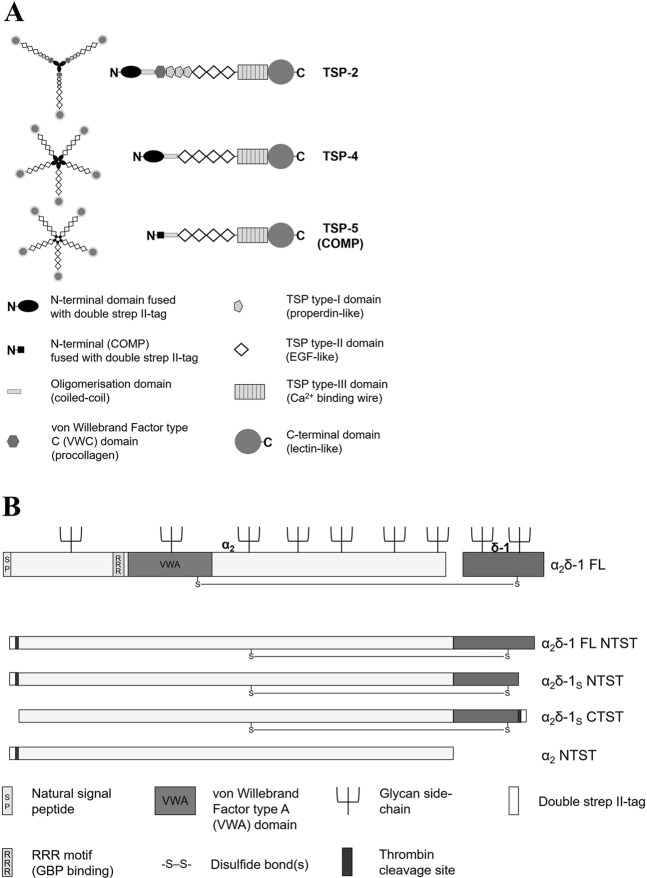
Thrombospondins (TSPs) form a family of five large oligomeric extracellular matrix glycoproteins that are expressed by numerous cell types, playing important roles in cellular migration, attachment and cytoskeletal dynamics1,2. Several TSP isoforms have been shown to be involved in a variety of physiological and pathological processes, including regulation of angiogenesis, apoptosis and platelet aggregation3–5. TSPs can be subdivided into subgroups A (TSP-1 and 2) and B (TSP-3–5, with TSP-5 also referred to as cartilage oligomeric matrix protein (COMP)) based on their oligomerisation state (trimeric or pentameric, respectively) and domain structure (Fig. 1A). In neurons, astrocyte-secreted or recombinantly expressed TSP(s), particularly TSP-1, TSP-2 and TSP-4, were reported to promote the formation of excitatory synapses both in vitro and in vivo through interaction with the voltage-gated calcium channel subunit α2δ-16–10. The α2δ proteins (α2δ‐1–4) are auxiliary subunits of voltage-gated calcium channels CaV1 and CaV2, and were found to be encoded by four different genes11–13. Functions of these auxiliary subunits include the modulation of trafficking, expression in the plasma membrane14–17, and biophysical properties of the channels15,17–19. Importantly, α2δ-1 acts as a specific binding site for gabapentinoid drugs20,21, mediating their analgesic effect in neuropathic pain21–23. Furthermore, studies using different animal models of neuropathic pain indicated the involvement of α2δ-1 in pain development, with nerve injuries leading to up-regulation of α2δ‐1 in both dorsal root ganglion (DRGs) and spinal dorsal horn neurons24–27 as well as to an increase of miniature excitatory post-synaptic current (mEPSC) frequency in the latter neurons22,25,26,28–30. Similarly, injury-induced TSP-4 is reported to mediate central sensitisation and neuropathic pain states8,9,31–37. This effect was recently shown to be mediated by activation of a TSP-4/α2δ‐1-dependent pathway which requires a direct molecular interaction between the two proteins9,34. Furthermore, the presence of TSP-4 was shown to modestly but significantly reduce the binding affinity of 3H-gabapentin (3H-GBP) towards α2δ‐1 in membrane preparations from TSP-4/α2δ‐1 co-transfected cells38. Taken together, the TSP-4/α2δ‐1 protein-protein interaction seems to be of potential translational importance and thus may serve as a novel target for developing a new class of analgesics against neuropathic pain.
Figure 1.
Schematic presentation of the structures of the recombinant proteins generated in this study. (A) Domain structure and oligomerisation state of the generated recombinant full-length TSP-2 (trimer), TSP-4 and COMP (pentamers). Schematic representation adapted by permission from Springer Nature, Cell Mol Life Sci, Structures of thrombospondins, Carlson, C. B., Lawler, J. & Mosher, D. F., Copyright (2008)39. All recombinant TSPs have been expressed with an N-terminal double strep II-tag and contain glycan side-chains which are not shown for reasons of clarity. (B) Structure of α2δ-1 FL protein (adapted from Cell 139, Eroglu, Ç. et al., Gabapentin receptor αδ2δ-1 is a neuronal thrombospondin receptor responsible for excitatory CNS synaptogenesis, 380–392, Copyright (2009), with permission from Elsevier7) and simplified depiction of the derived non-proteolytically processed α2δ-1 mutants generated in this study. The RRR motif, the von Willebrand Factor type A domain, and the glycan side-chains are not shown in the α2δ-1 mutants for reasons of clarity.
The aim of the present study is to investigate the biochemical characteristics of the direct molecular interaction between TSPs and α2δ‐1, addressing the question whether α2δ‐1 binding is specific to TSP-4 or redundant among other TSPs. GBP has been shown so far to inhibit the interaction of α2δ‐1 with a truncated form of TSP-2 in co-immunoprecipitation experiments7 as well as functionally by inhibiting synaptogenesis7,8,10, neuron sensitisation and behavioural hypersensitivity induced by TSP-2, its truncated fragment and/or TSP-49,34,35. Thus, we examined whether the direct TSP/α2δ‐1 interaction can be inhibited by GBP on a molecular level as well. We therefore generated purified recombinant forms of three full-length TSPs (TSP-2, TSP-4 and COMP) as well as soluble forms of α2δ‐1 subunit (α2δ‐1S), that shows GBP binding affinity similar to that of wild-type α2δ-1, and the α2 peptide chain of α2δ‐1 (Fig. 1). Both the interaction of these recombinant TSPs with α2δ‐1 and the possible inhibition by GBP were examined in a solid-phase ELISA-style ligand binding assay, with the capability of soluble α2δ‐1 to interact with GBP being proven by a newly developed surface plasmon resonance (SPR)-based binding assay. In order to demonstrate the characteristics of the direct TSP-4/α2δ‐1 interaction in an environment similar to that of native cells, we attempted to visualise the interaction of fluorescently labelled TSP-4 with membrane-localised full-length (FL) α2δ‐1 in a cell-based system.
Results
Biochemical characteristics of recombinant purified proteins expressed in HEK293-EBNA cells
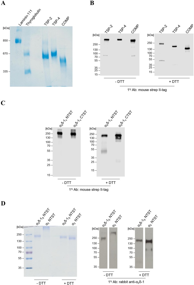
To investigate the direct binding of different TSPs to α2δ-1, three full-length recombinant TSPs, namely, the trimeric TSP-2, the pentameric proteins TSP-4 and COMP, and a soluble C-terminal deletion mutant of α2δ-1 carrying an N-terminal (α2δ-1S NTST) or a C-terminal (α2δ-1S CTST) double strep II-tag (Fig. 1) were generated in a eukaryotic expression system. Coomassie stained gels and immunoblots of the purified proteins confirmed their purity, identity and integrity (Fig. 2; Supplementary Fig. S1). As expected for TSPs, the intact proteins showed high molecular weight bands with approximate apparent molecular weights (Mr) in the range ~500–670 kDa when compared to laminin-111 and thyroglobulin as marker proteins (Fig. 2A). The oligomerisation patterns of these high molecular weight TSPs (i.e. pentamers for TSP-4 and COMP and trimer for TSP-2) were confirmed by comparing immunoblots in the absence and presence of the reducing reagent DTT (Fig. 2B; Supplementary Fig. S1a). In the latter case, DTT reduces the interchain disulphide bonds within the oligomerisation domains of the analysed TSPs39 and major bands of monomeric proteins with Mr of TSP-2 (~240 kDa), TSP-4 (~160 kDa), and COMP (~130 kDa) were observed (Fig. 2B, +DTT; Supplementary Fig. S1a, +DTT).
Figure 2.
The generated recombinant TSPs and α2δ-1S variants show high degree of purity and integrity in Coomassie staining and western blot analyses. (A,D left) Representative Coomassie-stained gels and (B,C and D right) immunoblots of three full-length TSP proteins, all carrying an N-terminal double strep II-tag: TSP-2, TSP-4, and COMP (A,B); α2δ-1S variants carrying either an N-terminal (α2δ-1S NTST) or a C-terminal (α2δ-1S CTST) double strep II-tag (C); α2δ-1S NTST and α2 peptide chain carrying an N-terminal double strep II-tag, α2 NTST (D). Proteins were separated under non-reducing (−DTT) or reducing conditions (+DTT) on 4–15% (B), 10% (C), and 7% (D) polyacrylamide gels, respectively, while in (A) proteins were separated on 0.5% agarose (w/v)/3% polyacrylamide (w/v) composite gels without prior DTT treatment. Proteins were either stained with colloidal Coomassie stain (A,D left) or detected with the following primary antibodies after blotting: mouse anti-strep II-tag (B,C) or rabbit anti-α2δ-1 (D right). Secondary antibodies included the polyclonal rabbit anti-mouse IgG (B,C) and swine anti-rabbit IgG (D right), both conjugated with horseradish peroxidase (see Supplementary Table S2 for further information). In all gels the molecular weight standard (in kDa) indicated on the left was PageRuler Plus Prestained Protein Ladder (Thermo Fisher Scientific) except for (A) in which both thyroglobulin (Sigma) and recombinant laminin-111 (kind gift from Prof. Dr. Monique Aumailley, Institute for Biochemistry II, Centre for Biochemistry, Medical Faculty, University of Cologne) were used.
Similar analysis was performed for the generated α2δ-1S NTST and the respective C-terminally tagged α2δ-1 variant, α2δ-1S CTST, showing single but smeared bands at approximate Mr ~200 kDa under non-reducing conditions (Fig. 2C, −DTT; Supplementary Fig. S1b, −DTT) and appear as distinct bands at approximate Mr ~180 kDa under reducing conditions (Fig. 2C, +DTT; Supplementary Fig. S1b, +DTT). So far, there is no comprehensive explanation for this gel band shift in the presence of DTT. However, it cannot be attributed to the reductive cleavage of the interchain disulphide bridge between α2 and δ-1 followed by loss of the smaller δ-1 chain. This conclusion arises from the observation that α2δ-1S bearing the C-terminal double strep II-tag (α2δ-1S CTST) is still detectable in immunoblots probed with strep II-tag antibody following DTT treatment. In agreement with this result, mass spectra of α2δ-1S NTST recorded with and without DTT pre-treatment showed almost identical molecular ion peaks ([M + H]+; Table 1, Supplementary Fig. S2d,e). This observation confirms the results by Brown and Gee40 who first described a similar soluble mutant of the porcine α2δ-1 orthologue which retains high affinity for 3H-GBP. Although uncleaved α2δ-1 may not represent a functional form as a subunit of the CaV channels and can inhibit native calcium currents in mammalian neurons41, the TSP/α2δ-1 pathway is thought to be at least partially independent of the roles of α2δ-1 as a CaV channel subunit7,10. Therefore, the recombinant uncleaved α2δ-1S variant used in this study should be suitable for the purpose of investigating TSP binding biochemically. Notably, we observed a minor band in the immunoblots of α2δ-1S CTST at Mr ~25 kDa upon DTT treatment and detection with strep II-tag antibody (Fig. 2C, +DTT) which is most likely attributed to cleaved strep-tagged δ-1S chain. This indicates the presence of a small fraction of the generated purified α2δ-1S in a proteolytically cleaved form.
Table 1.
Molecular masses of recombinant TSPs and α2δ-1S NTST obtained by MALDI-TOF MS.
| Protein | m/zcalc [M + H]+ | m/zexp [M + H]+ |
|---|---|---|
| TSP-2 (monomer) | 132,137 | 146,958 |
| TSP-4 (monomer) | 107,409 | 109,351 |
| COMP (monomer) | 84,714 | 88,463; 87,178 |
| α2δ-1S NTST (−DTT) | 123,000 | 152,236 |
| α2δ-1S NTST (+DTT) | 112,130* | 152,258 |
The m/zcalc [M + H]+ values for all proteins (in Da) were calculated based on their amino acid sequences using ExPASy Compute pI/Mw online tool, while the m/zexp [M + H]+ values (in Da) were obtained from the MALDI-TOF MS spectra of the respective proteins.
* m/zcalc [M + H]+ of α2 NTST was calculated for the expected product of the DTT-mediated reduction of the disulphide bond between α2 and δ-1S in α2δ-1S NTST.
In addition to the α2δ-1S variants generated, the α2 peptide chain (α2 NTST) was recombinantly produced in a similar way (Fig. 1B). Expression and purification of this fragment as well as analyses by SDS-PAGE and immunoblotting (Fig. 2D) were carried out as described above. Here, we observed the formation of a high molecular weight product under non-reducing conditions which dissociated into the monomeric form after DTT treatment (approximate Mr ~ 170 kDa, Fig. 2D) which points to the formation of interchain disulphide bonds in the absence of reducing agents (see also Discussion section below).
Notably, all recombinant proteins generated in this study that had been analysed by SDS-PAGE and Western Blot showed protein bands at remarkably higher Mr than expected from their amino acid sequences. It is known that the electrophoretic mobility of proteins can be greatly influenced by the extent of post-translational modifications (e.g. glycosylation) of the protein where the glycan chains do not bind SDS leading in many cases to decreased mobility, and increased Mr, of the glycoprotein analysed by SDS-PAGE42. In addition, sample treatment prior to loading onto the gel (e.g. heating at 95 °C with DTT) represents a possible source of abnormal protein migration on the gels through its impact on the structure of the analysed protein43. Therefore, further analysis was carried out to determine the accurate molecular masses of the recombinant purified proteins using MALDI-TOF mass spectrometry. The results are shown in Table 1 and Supplementary Fig. S2. The molecular masses of TSP-4 and COMP, both in monomeric form, were found to be only slightly higher (1–3 kDa) than the theoretical masses calculated on the basis of each protein’s amino acid sequence, indicating minor post-translational modifications, e.g. glycosylation, of these proteins. In case of TSP-2 and α2δ-1S NTST, the experimentally determined masses were about 15 and 30 kDa, respectively, larger than the theoretical ones, which is likely attributed to heavy glycosylation, as shown previously for α2δ-1 in the work of Kadurin et al.44 (Table 1, Supplementary Fig. S2a,d,e).
Characterisation of the interaction of TSPs with α2δ-1S using an ELISA-style ligand binding assay
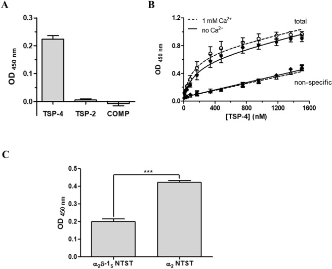
First, the purified recombinant full-length TSPs (soluble) were examined for their direct interaction with immobilised α2δ-1S NTST in an ELISA-style ligand binding assay, that was validated using the model interaction of COMP and matrilin-3 proteins45 (Supplementary Fig. S4). Preliminary experiments with TSP concentrations up to ~500 nM had demonstrated that out of the three TSPs generated in this study, only TSP-4 was able to directly interact with α2δ-1S NTST (data not shown). To rule out the possibility of a very low binding affinity of TSP-2 and COMP towards α2δ-1, we finally utilised a comparably high concentration (1,000 nM) of all TSP proteins in the same ELISA which confirmed our preliminary data (Fig. 3A). Therefore, we decided to focus on the interaction of TSP-4 with α2δ-1S and study it more in detail. Notably, we detected comparable binding signals of TSP-4 to either α2δ-1S NTST or α2δ-1S CTST variants in a preliminary experiment (data not shown) and hence we chose to proceed with one of the two variants, namely, α2δ-1S NTST in our binding assays. Titration of immobilised α2δ-1S NTST with increasing concentrations of TSP-4 (11-1,505 nM) showed saturable binding with an apparent KD value of about 200 nM. Since Ca2+ binding is associated with major conformational changes and structural rearrangements of both TSPs46,47 and the metal ion-dependent adhesion site (MIDAS) motif of α2δ-117,48, we investigated the effect of Ca2+ on TSP-4/α2δ-1S NTST interaction. Our data show that the apparent KD value was slightly, but not significantly, decreased in presence of 1 mM Ca2+ (Table 2, Fig. 3B). Similarly, the maximum binding value (Bmax, unitless) showed a small, statistically non-significant increase in presence of 1 mM Ca2+ (Table 2, Fig. 3B). These results indicate that the TSP-4/α2δ-1S NTST interaction is not sensitive to Ca2+ changes. Next, the ability of TSP-4 to directly interact with the α2 fragment of α2δ-1 was analysed. Here, we observed a two-fold, statistically significant increase in the binding signal of a single concentration of TSP-4 (1,000 nM) when using immobilised α2 NTST instead of α2δ-1S NTST (Fig. 3C). These results indicate the localisation of the TSP-4 binding site(s) within the α2 region of α2δ-1 in agreement with data obtained by Eroglu et al.7. The enhanced binding signal of TSP-4 to α2 NTST is suggestive of a more favourable conformation of α2 NTST which is more accessible for TSP-4 binding, as compared to the non-proteolytically cleaved α2δ-1S NTST.
Figure 3.
The direct binding to α2δ-1S NTST is TSP-4 specific in an ELISA-style ligand binding assay. α2δ-1S NTST (20 µg/ml) was coated onto 96-well plates and incubated with either (A) TSP-2, TSP-4 or COMP (1,000 nM), or (B) increasing concentrations of TSP-4 (11–1,505 nM). Shown are data for total (circles) and non-specific (triangles, rhombi) binding in the absence (full symbols) and presence (open symbols) of 1 mM Ca2+. (C) Soluble α2 NTST or α2δ-1S NTST (10 µg/ml) were coated onto 96-well plates and incubated with TSP-4 (1,000 nM). Binding assays (A,C) were carried out in the presence of 1 mM Ca2+ and bound proteins were detected with the corresponding TSP-specific antibody/antiserum (see Supplementary Table S2). Specific binding in (A,C) was calculated by subtracting OD values of non-specific binding from those of total binding. Data of total and non-specific binding were used to calculate KD and Bmax in (B), with the linear dependence of the non-specific signal on the TSP-4 concentration ensuring the absence of perturbations of the assay system. Data represent mean values ± SEM of 3 independent measurements performed in duplicates or triplicates. Statistical analysis in (C) was done using unpaired two-tailed Student’s t-test (***P = 0.0003).
Table 2.
Binding parameters (KD, Bmax) for the interaction of TSP-4 with α2δ-1S NTST.
| Assay buffer | KD (nM) | Bmax |
|---|---|---|
| TBS | 198 ± 49 | 0.579 ± 0.066 |
| TBS + 1 mM Ca2+ | 153 ± 46 | 0.677 ± 0.054 |
Data represent mean values ± SEM of 3 independent experiments, each performed in duplicate. Statistical analysis by an unpaired two-tailed Student’s t-test showed no significant difference for KD (apparent dissociation constant, P = 0.5381) and Bmax (maximum binding, unitless, P = 0.3138) obtained in the absence and presence of Ca2+.
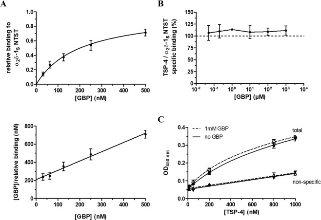
To investigate the effect of the known α2δ-1 ligand GBP20 on the observed TSP-4/α2δ-1S interaction, we checked for the ability of the recombinantly expressed mutant α2δ-1S NTST to bind GBP with high affinity (KD = 219 nM) using a label-free surface plasmon resonance (SPR) assay (Fig. 4A, Supplementary Fig. S3). Our results are in agreement with the binding data for 3H-GBP to membrane preparations of heterologously expressed human α2δ-1 (KD range of 140–175 nM)38. Various GBP concentrations (0.05–1,000 µM) were used in the TSP-4 /α2δ-1S NTST binding assay to interfere with this protein-protein interaction but did not show inhibitory effects (Fig. 4B). To rule out the possibility that the GBP binding pocket might not be accessible after immobilisation of α2δ-1S NTST on the ELISA microplate, we had coated the wells with α2δ-1S NTST after pre-incubating the protein with GBP. In addition, GBP had been supplemented to the liquid phase during blocking and incubation with TSP-4 ensuring availability of a sufficient number of GBP molecules to α2δ-1S NTST and preventing dissociation of bound GBP during the experiment. In a further experiment, the binding of various concentrations of TSP-4 (12.5–1,000 nM) to α2δ-1S NTST was not affected by the highest GBP concentration (1,000 µM) investigated (Fig. 4C). Together, these data suggest that GBP alone is not sufficient to disrupt the interaction of TSP-4 with α2δ-1 on a molecular level.
Figure 4.
GBP does not directly interfere with the binding of TSP-4 to α2δ-1S NTST in an ELISA-style ligand binding assay. (A) Surface plasmon resonance (SPR) measurements of the binding of GBP to recombinant α2δ-1S NTST. The protein (10–15 µg/ml) was directly immobilised to CM5 sensor chips and GBP (31.25–500 nM) in PBS buffer, pH 7.4 containing 0.05% Tween 20 was passed over the chip at a flow rate of 30 μl/min. Shown are data of the relative GBP binding to α2δ-1S NTST (Top) obtained from single cycle kinetics protocol (mean values ± SEM of 4 independent experiments, Fig. S3). Each experiment was analysed by non-linear regression according to the equation RU/RUmax = [GBP]/(KD + [GBP]), where the ratio of the binding response and the maximum binding response, RU/RUmax, represents the relative binding at a given GBP concentration, [GBP], and KD is the dissociation constant of the two interaction partners. Data analysis yielded a value of KD = 219 ± 47 nM (mean value ± SEM, n = 4), with the linear shape of the Hanes-Woolf transformation (Bottom) showing equimolar binding of the two interaction partners. For the ELISA-style assay, the α2δ-1S NTST (10 µg/ml) protein was coated onto 96-well plates and incubated with either (B) TSP-4 (1,000 nM) in the absence and presence of increasing concentrations of GBP (0.05–1,000 µM), or (C) increasing concentrations of TSP-4 (12.5–1,000 nM) in the absence (full symbols) and presence (open symbols) of GBP (1,000 µM). The assay was carried out in the presence of 2 mM Mg2+ and bound TSP-4 was detected with TSP-4-specific antiserum. Specific binding was calculated by subtracting OD values of non-specific binding (triangles) from those of total binding (circles). Data in (B) and (C) represent mean values ± SEM of 2 to 3 independent experiments, each performed in duplicate. In (B) the OD values for specific binding of TSP-4 in the presence of GBP (0.05–1,000 µM) were normalised to those in the absence of GBP.
Fluorescent A555-TSP-4 does not bind to membrane-bound α2δ-1 in a cell-based binding assay
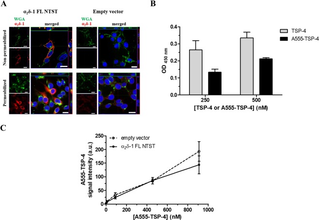
To examine the interaction between TSP-4 and full-length membrane-bound α2δ-1 in a cellular environment, HEK293-EBNA cells were transfected with either empty vector or vector encoding α2δ-1 FL NTST. Immunocytochemical analysis of cells transfected with α2δ-1 FL NTST revealed a marked increase in α2δ-1 immunoreactivity, confirming the over-expression of heterologous α2δ-1 (Fig. 5A, left column).
Figure 5.
Fluorescent A555-TSP-4 protein does not bind to membrane-bound α2δ-1 in HEK293-EBNA cells. (A) Representative confocal images of the immunocytochemical detection of membrane-localised α2δ-1 FL NTST over-expressed in HEK293-EBNA cells. Cells were transfected with either empty vector (right column) or vector encoding full-length α2δ-1 containing an N-terminal strep II-tag (α2δ-1 FL NTST, left column). Cells were stained either without permeabilisation (upper row) or after membrane permeabilisation (lower row). Signals of WGA conjugated with Alexa Fluor 633 (green) and α2δ-1 (red) are shown individually in the small images; merged signals are shown in the large images. DAPI was used to visualise the nucleus (blue). Images show top view as well as upper-side (green box) and right-side (red box) views of a single slice of scanning near the middle of cells. Scale bar is 20 µm for all images. (B) Binding of A555-TSP-4 or TSP-4 to α2δ-1S NTST analysed by an ELISA-style ligand binding assay. α2δ-1S NTST (20 µg/ml) was coated onto 96-well plates and incubated with either TSP-4 or A555-TSP-4 at two different concentrations (250 and 500 nM) for each protein. Specific binding was calculated by subtracting OD values of non-specific binding from those of total binding. Data represent mean ± SEM of 2 independent measurements, each performed in duplicate. OD values for specific binding of TSP-4 and A555-TSP-4 were subjected to unpaired Student’s t-test. No significant difference was found (P > 0.05) for each of the two concentrations of both TSP-4 species used. (C) Cells transfected with either empty vector or vector encoding α2δ-1 FL NTST were incubated with increasing concentrations of A555-TSP-4 (final concentration: 9–909 nM) in 96-well imaging plates. Control wells received the same volume of dilution medium (40 µl) without A555-TSP-4. Average A555-TSP-4 fluorescent signal intensities from wells containing cells transfected with α2δ-1 FL NTST encoding vector or empty vector (±SEM of 3 independent experiments, each performed in triplicate) were plotted versus the A555-TSP-4 concentration.
In cells immunostained without permeabilisation, a high degree of co-localisation of α2δ-1 and wheat germ agglutinin (WGA) used for labelling glycoproteins or glycolipids of the outer leaflet of the plasma membrane49 were observed, assuring the localisation of heterologous α2δ-1 in the plasma membrane (Fig. 5A, upper left image). The transfected cells were incubated with increasing concentrations of fluorescently labelled A555-TSP-4 (9–909 nM), the binding of which to α2δ-1S NTST was found to be non-significantly lower than that of unlabelled TSP-4 in ELISA-style assay (Fig. 5B). Images of the stained cells were acquired using a High Content Screening microscope and the average A555-TSP-4 signal intensity was determined for each well. In addition, α2δ-1 FL NTST was visualised by immunostaining to analyse the co-localisation of A555-TSP-4 with α2δ-1 FL NTST. Results show low overall A555-TSP-4 signal intensity which increases upon increasing the concentrations of the fluorescent protein. However, no difference was observed when comparing α2δ-1 FL NTST- and empty vector-transfected cells (Fig. 5C). Furthermore, no co-localisation of A555-TSP-4 and α2δ-1 FL NTST signals was detected in these experiments (Supplementary Fig. S5). These results are in accordance with recently published data by Lana et al.38 showing no TSP-4 co-localisation with α2δ-1 on the cell surface of tsA-201 cells co-expressing both proteins or in mixed populations of cells transfected separately with either α2δ-1 or TSP-4.
Discussion
All TSPs, i.e. TSPs 1–4 and COMP, were previously identified as synaptogenic proteins which, together with other astrocyte-derived factors, help to promote the formation of functional excitatory synapses in the CNS6,7. The α2δ-1 protein was demonstrated to be functionally involved in TSP-induced synaptogenesis by means of synaptic assays in retinal ganglion cells (RGCs)7, DRG/spinal cord primary neuron co-culture8,9, purified cortical neurons10 as well as in dorsal spinal cord of mice34. Biochemically, α2δ-1 was reported to interact in co-immunoprecipitation experiments with TSP-1, TSP-2 and TSP-4 from rat cerebral cortex7 as well as with TSP-4 from rodent spinal cord34. Similarly, a TSP-2 fragment containing all three EGF-like repeats, the calcium-binding repeats, and the C-terminal globular domain was co-purified with full-length α2δ-1 or its protein-binding VWA domain after co-expression in HEK293 cells7. Recently, Park et al.9,34 demonstrated for the first time a direct molecular interaction between α2δ-1 and recombinant full-length TSP-4 or its fragments containing EGF-like or coiled coil domains. In the present study, we investigated the biochemical properties of the direct TSP-4/α2δ-1 interaction. Furthermore, it was of importance to know whether other members of the TSP protein family are also able to directly bind to α2δ-1 in an analogous manner to that of TSP-4. Our data demonstrated that only full-length TSP-4, but not TSP-2 or COMP, is able to directly interact with immobilised soluble α2δ-1 variant (α2δ-1S NTST) in an ELISA-style ligand binding assay (Fig. 3A), indicating the specificity of this protein-protein interaction. This observation is in direct contrast to that of Eroglu et al.7 (see above). Nevertheless, TSP-4 is remarkably the only isoform of TSP proteins reported so far to be implicated in neuropathic and joint-mediated chronic pain in rodents along with neuronal α2δ-18,9,31,34,35. During the processes resulting in such pain, both TSP-4 and α2δ-1 are up-regulated on the protein level and temporally correlate with the development of behavioural hypersensitivity in the respective animal models (for review see ref.50). In contrast, TSP-1/-2, though previously shown to be up-regulated after ischemic brain injury in rodents4,51 and promoting the subsequent synaptic recovery51, are not dysregulated on the protein level in dorsal spinal cord after spinal nerve ligation in mice, even when behavioural hypersensitivity was evident in these animals. This observation led Kim et al.31 to rule out the possibility of the involvement of these two astrocyte-secreted proteins in mediating TSP/α2δ-1-induced neuropathic pain. With regards to COMP, it has been reported to be expressed in several tissues including skeletal muscle, tendon, and cartilage. In the latter tissue, COMP is known to be mainly involved in chondrocyte differentiation, attachment, and cartilage extracellular matrix assembly52–55, with mutations in COMP being associated with pseudoachondroplasia56,57. COMP in skeletal muscle, tendons, and perichondrium can be theoretically in contact with nerve terminals containing α2δ-1. In addition, COMP was shown to have synaptogenic potential in RGCs (as discussed) and shares a high degree of both overall sequence identity (~70%) and structural similarity (Fig. 1A) with TSP-4. That is why we investigated COMP as a potential interaction partner of α2δ-1 in our binding studies. Nevertheless, COMP is, in contrast to TSP-4, neither abundant in neurons and astrocytes nor it is known to be dysregulated in neuropathic pain states. The fact that, beside TSP-4, all other TSPs were previously found to induce synaptogenesis through a mechanism involving neuronal α2δ-17 may refer to other cellular factors or scaffold proteins required to mediate their interaction with α2δ-1 indirectly. Furthermore, it is tempting to speculate that TSP-induced synaptogenesis might be of little relevance to neuropathic pain development since, as previously mentioned, TSP-4 is the only member of TSP family found to be up-regulated in dorsal spinal cord following nerve injury. Indeed, a recent study shows that an enhanced presynaptic NMDA receptor activity, rather than synaptogenesis, is responsible for maintaining the increased synaptic excitatory transmission in dorsal spinal cord leading to chronic pain states following nerve injury in mice58.
In further experiments, we observed a significantly increased binding of TSP-4 to α2 NTST when compared to α2δ-1S NTST (Fig. 3C), confirming previous data by Eroglu et al.7 demonstrating the TSP-4 binding site to be localised within the α2 region of α2δ-1 (VWA domain). The observed enhancement in binding towards α2 might be attributed to more exposed TSP-4 binding motif(s) in the immobilised α2 NTST compared to the non-proteolytically processed α2δ-1S NTST. This result is in agreement with recent findings from Lana et al.38 where wild-type α2δ-1 was very weakly co-immunoprecipitated with TSP-4, but no co-immunoprecipitation of the mutant α2δ-1 (MIDASAAA) with TSP-4 was detected in lysates of co-transfected tsA-201 cells. In addition, the binding to α2 NTST seems again to be TSP-4-specific since negligible binding signals were detected when equimolar concentrations of either COMP or a truncated TSP-4 fragment were utilized in a pilot experiment (data not shown). Although our results are supported by reported data, we cannot rule out the possibility that the enhanced TSP-4 binding signal is due to improperly expressed α2 NTST since unpaired cysteine residues, normally involved in the formation of disulphide bridges with other cysteine residues in the deleted regions of wild-type α2δ-159, become available. It is worth mentioning here that a very high tendency for multimerisation was observed for a recombinant VWA domain of α2δ-1 generated in this study (data not shown) due to the formation of intermolecular disulphide bonds. Therefore, it might be appropriate to consider the expression of α2 and VWA fragments in which the unpaired cysteines are replaced by other isosteric residues (e.g. serine) for future binding studies.
One of the reported small molecules capable of interfering with the TSP/α2δ-1 interaction is GBP, an approved analgesic against neuropathic pain60–62 and a known ligand of α2δ-120. Biochemically, co-immunoprecipitation experiments showed that the interaction between a truncated TSP-2 fragment and α2δ-1 FLAG in a co-culture of two populations of HEK293 cells was diminished in the presence of GBP7. In addition, as previously mentioned, TSP-4 modestly but significantly reduces the binding affinity of 3H-GBP to α2δ-1, suggesting rather an allosteric than a pure competitive mode of inhibition38. Furthermore, in vivo data revealed the ability of GBP to block TSP-4-induced neuronal sensitisation and behavioural hypersensitivity as well as changes in Ca2+ currents and intracellular Ca2+ transients after injuries to peripheral nerves or facet-joint in rodents8,9,34,35,63. Similarly, several studies in neuropathic pain models demonstrated the ability of GBP to inhibit α2δ-1-induced26 or TSP-induced7,8,34,35 synaptogenesis. Most recently, GBP was also shown to inhibit TSP-2-induced synapse formation in purified culture of cortical neurons10. Despite the multidimensional evidence of GBP interference with TSP/α2δ-1 interaction, a direct GBP inhibition of this interaction on the molecular level has never been investigated before, to our knowledge. In the current study, we did not observe any inhibition of the direct TSP-4/α2δ-1S NTST interaction in the presence of increasing concentrations (up to 1 mM) of GBP (Fig. 4B). Furthermore, the highest GBP concentration used (1 mM) did not shift the TSP-4/α2δ-1S NTST binding curve (Fig. 4C). Although the utilised α2δ-1S NTST was mostly expressed as uncleaved form of the protein (in agreement with the original work describing a similar porcine α2δ-1 mutant40), we were able to demonstrate the ability of this α2δ-1S mutant to retain high affinity for GBP (Fig. 4A). For this purpose, a newly developed SPR-based binding assay suitable for detecting and quantifying the binding of small molecules to immobilized recombinant α2δ-1S was used. This SPR assay has the advantage of being radiolabel-free and can easily be used to determine the binding kinetics unlike the previously used 3H-GBP binding assay38,40,64,65. Taken together, our data confirmed that the proteolytic cleavage of α2δ-1 is not crucial for the formation of the GBP binding pocket40. The complete lack of GBP inhibition towards the interaction of purified TSP-4 with α2δ-1S NTST raises questions regarding the exact mechanism by which GBP can block the above-mentioned TSP-induced changes. It is possible that other unknown factors in the cellular environment are essential for GBP to interfere with α2δ-1/TSP-4 interaction and thereby mediating the known GBP inhibitory effects. Another possible explanation based on the recent findings of Chen et al.58,66 is that the α2δ-1/NMDA receptor complex, rather than α2δ-1/TSP-4 binding, represents the molecular target of gabapentinoid drugs to alleviate neuropathic pain.
Our efforts were as well focused on the investigation of the TSP-4/α2δ-1 interaction in a cellular system to get closer to the physiological/pathological situation in the CNS. We over-expressed full-length α2δ-1 in HEK293-EBNA cells and demonstrated both its intracellular and plasma membrane localisation in transfected cells (Fig. 5A). Treatment with increasing concentrations (up to 909 nM) of fluorescently labelled A555-TSP-4, however, showed no differences in binding of the protein to α2δ-1 overexpressing cells when compared to control cells (Fig. 5C, Supplementary Fig. S5). Furthermore, fluorescent signals of A555-TSP-4 did not co-localise with those of immunostained α2δ-1 on the cells (Supplementary Fig. S5). The observed loss of binding cannot be attributed to an impairment of the interaction of the two proteins by the fluorescent label of TSP-4 since the fluorescent protein was generated with a minimal dye-to-protein molar ratio and showing substantial α2δ-1S NTST binding in the ELISA-style assay (Fig. 5B). A possible explanation could instead be arising from the weak interaction of the two proteins under the conditions of the cell-based assay, unlike the ELISA-style assay. This means that very high local concentrations of TSP-4 in the proximity of cell-surface α2δ-1 would be required to enable the detection of their interaction by simulating the pathological situation (e.g. dramatic up-regulation following nerve injury). This could not, however, be achieved with the range of A555-TSP-4 concentrations (up to 909 nM) used in the assay. In our experiments, we over-expressed α2δ-1 in HEK293-EBNA cells without co-expression of α1 subunit, which interacts intracellularly with α2δ-1 before trafficking of the complex to the cell surface67. However, we assume that the lack of α1 subunit did not hamper the putative binding of TSP-4 to α2δ-1 on the cell surface. This assumption is based on recent data showing the ability of wild-type α2δ-1, expressed in HEK293 cells without co-expression of α1 subunits, to be efficiently transported to the cell surface and thereby become accessible to extracellular ligands like TSP10. Functionally, this α2δ-1 over-expression construct alone was able to rescue synapses in cortical organotypic slices from α2δ-1 knockout mice. This effect is found to be mediated through activation of the small Rho GTPase Ras-related C3 botulinum toxin substrate 1 (Rac1) and is independent of α1 subunits of the postsynaptic L-type calcium channels CaV1.2 and CaV 1.310.
The α2δ subunits are thought to promote membrane trafficking of the pore subunits of voltage-gated calcium channels17 and α2δ-1-driven allodynia in mice can be reversed by blockers of voltage-gated calcium channels like ω-conotoxin GVIA68. However, other findings suggest that the maladaptive changes contributing to chronic pain in rodents following nerve injuries and resulting from the interaction of dysregulated TSP-4 with α2δ-1 are partially independent of the role of the latter protein in regulating voltage-gated calcium channels’ trafficking and function50.
As previously mentioned, our data align with those of Lana et al.38 who reported no interaction of secreted TSP-4 with membrane-localised α2δ-1 on tsA-201 cells when subjected to immunocytochemical analysis. On the other hand, the same study showed weak intracellular interaction of both proteins in co-immunoprecipitation experiments from cells over-expressing both proteins38. It has therefore been postulated that the weak TSP-4/α2δ-1 interaction may occur in an intracellular compartment rather than on the cell surface38,69. This postulation is in contrast to the previous data showing synaptogenic effect of secreted TSPs which is mediated by neuronal α2δ-1 thought to be located either pre-8 or post-7,10 synaptically. To our knowledge, there are no data so far showing the co-localisation of TSP(-4) and α2δ-1 in neuronal cell cultures or spinal cord tissue where TSP-induced changes (e.g. synaptogenesis) were demonstrated. To reveal the exact cellular localisation of TSP-4/α2δ-1 interaction it would be very helpful to simultaneously analyse co-immunostained TSP-4 and α2δ-1 proteins in cultures of neurons utilized in in vitro synapse assays6,7,10.
In summary, our results provide substantial in vitro biochemical evidence for a direct and specific Ca2+-insensitive TSP-4/α2δ-1 interaction which is rather weak. Importantly, GBP does not inhibit this interaction on a molecular level, indicating the possible involvement of other unknown factors or targets in mediating GBP inhibitory effects in neuropathic pain.
We, therefore, need to understand the exact and complete molecular mechanism of the TSP/α2δ‐1 interaction to really be able to design appropriate small molecule modulators - rather than being left to use and optimize the enigmatic properties of the serendipitously discovered gabapentinoid action.
Materials and Methods
Cloning, expression and purification of recombinant proteins
The plasmids encoding full-length murine TSP-2 (TSP-2, accession no. of the translated protein product AAH53702.1), full-length rat COMP (COMP, accession no. of the translated protein product EDL90681.1), and full-length rat TSP-4 (TSP-4, accession no. of the translated protein product NP_058829.1) were generated as described earlier for COMP70. The cDNAs encoding human α2δ‐1 soluble variant40 with an N-terminal or a C-terminal double strep II-tag (α2δ‐1S NTST and α2δ‐1S CTST, respectively) and its α2 peptide chain with an N-terminal double strep II-tag (α2 NTST) were amplified following the same protocol from a pIRESpuro3-α2δ‐1 vector, originally generated from human α2δ‐1 FL cDNA71 (α2δ‐1, accession no. of translated protein product XP_005250627.1). All PCR products were lacking the natural signal peptide sequences and harboured a NheI restriction site at the 5′-end and a XhoI or BamHI site at the 3′-end. After digestion with NheI and either XhoI or BamHI, the amplified cDNAs were cloned into the modified episomal expression vector pCEP-Pu-double strep II-tag (N- or C-terminal)72 in-frame with the 5′ sequence of the BM-40 signal peptide and confirmed by Sanger nucleotide sequencing. Here, the sequences of the double strep II-tag and the thrombin cleavage site were directly located either at the 5′- or the 3′-end of the inserted cDNA sequences.
The recombinant plasmids carrying cDNAs encoding full-length proteins or truncated fragments were transfected into human embryonic kidney 293/Epstein-Barr virus nuclear antigen cells (HEK293-EBNA, Invitrogen) using TurboFect transfection reagent (Thermo Fischer Scientific). Transfected cells were cultured in DMEM medium supplemented with 10% FCS (Gibco), penicillin (1000 U/ml)/streptomycin (1000 µg/ml), and puromycin (3 µg/ml, Gold Biotechnology) for positive selection and were incubated at 37 °C/5% CO2 to allow growth to 100% confluency as adherent monolayers in cell culture triple-flasks (NuncTM, Thermo Fisher Scientific). Cell culture supernatants containing the secreted target proteins were collected and supplemented with phenylmethylsulfonyl fluoride (1 mM, Applichem) before passing over self-packed streptactin-sepharose columns (0.5 ml, IBA). The recombinant double strep II-tagged proteins were eluted with phosphate-buffered saline (PBS), pH 7.4 containing D-desthiobiotin (2.5 mM, Sigma), concentrated with Amicon ultra centrifugal filter units with molecular weight cutoff of 100 kDa for all recombinant proteins (Millipore), aliquoted after addition of glycerol (10% (v/v)) and stored at −80 °C until use.
Coomassie stained gels and immunoblotting analysis
For protein analysis by Coomassie staining, the purified TSPs were separated on 0.5% (w/v) agarose/3% (w/v) polyacrylamide composite gels73 without prior DTT treatment, while α2δ-1S NTST and α2 NTST variants were separated using conventional SDS-PAGE on 7% gels in presence and absence of DTT. Gels were stained overnight with colloidal Coomassie staining solution followed by destaining for 1–2 hours in a solution of ethanol (10% v/v) and o-phosphoric acid (2% v/v). All purified proteins were subjected to immunoblotting analysis by separating the proteins using SDS-PAGE under non-reducing and reducing conditions on 4–15% gradient gels for TSPs, 10% gels for α2δ‐1S NTST and α2δ‐1S CTST (before and after digestion with thrombin), and 7% gel for α2 NTST. Proteins were then transferred onto PVDF membranes by blotting overnight at 4 °C and blocked with 5% skimmed milk in Tris-buffered saline (TBS), pH 7.4 for 1 h at RT. The blocked membranes were incubated with antibodies recognizing the (double) strep II-tag or specific epitopes of the proteins for 1 h at RT, followed by incubation with the corresponding horseradish peroxidase-conjugated secondary antibodies (see list of antibodies used in Supplementary Table S2) for 1 h at RT. Protein bands were visualised using the Odyssey Fc Imaging system (LI-COR Biosciences).
Mass spectrometry
The masses of the purified recombinant α2δ‐1S NTST and TSPs were determined by MALDI-TOF mass spectrometry performed in the bioanalytical laboratory of the Centre for Molecular Medicine Cologne (CMMC) using a similar protocol to that described by Klatt et al.74. Briefly, TSPs and α2δ‐1S NTST were first incubated with DTT (10 mM) in PBS overnight at 4 °C with additional sample of α2δ‐1S NTST being treated similarly but without DTT. Next, samples were desalted with micro Zeba Spin desalting columns (Pierce Biotechnology, Thermo Fisher Scientific) equilibrated in trifluoroacetic acid (0.1% (v/v)) in ultrapure water. 5 µl of the eluate were mixed with 5 µl of a saturated solution of sinapinic acid (Bruker) in acetonitrile (50% (v/v)), trifluoroacetic acid (0.1% (v/v)), and 1 µl of the mixture was applied onto a MTP 384 polished steel target (Bruker). MALDI spectra were acquired with an Ultraflextreme MALDI-TOF/TOF instrument (Bruker) operated in linear mode. The monomer ([M + H]+, [M + 2 H]2+) and dimer ([2 M + H]+) peaks of bovine serum albumin (BSA, Applichem) were used for external calibration of the spectra. Analysis of the MS spectra was done with FlexAnalysis software (Bruker).
Fluorescence labelling of TSP-4
TSP-4 was labelled with Alexa Fluor 555 succinimidyl ester (Life Technologies) according to manufacturer’s instructions. Briefly, buffer exchange of protein samples to 100 mM NaHCO3, 100 mM NaCl (pH 8.3) was performed using Amicon ultra centrifugal filter unit (Millipore). Protein sample was then incubated with a five-fold amount of Alexa Fluor 555 succinimidyl ester (stock dissolved at 10 mg/ml in dry dimethylformamide) for 1 h at 25 °C followed by addition of 10% (v/v) hydroxylamine (1.5 M), pH 8.5, and incubation of the hydroxylamine-containing reaction mixture for 1 h to terminate the reaction. Free dye was then separated using gel filtration by loading the protein/dye reaction mixture onto a 15-ml self-packed Sephadex G-25 (Sigma) column and elution with PBS, pH 7.4. Fractions containing labelled protein eluted in the void volume were pooled, mixed with glycerol 10% (v/v) and stored at −80 °C until use.
ELISA-style ligand binding assay
Purified α2δ‐1S NTST (10 or 20 µg/ml) or α2 NTST (10 µg/ml) in coating buffer (TBS, pH 7.4 with or without 1 mM Ca2+ or with 2 mM Mg2+ for experiments with GBP) were coated overnight at RT onto 96-well plates (Nunc Maxisorb) to obtain 0.5–1 µg protein/well for determination of values of total binding. Values of non-specific binding were obtained by adding coating buffer to the wells and proceeding similarly. After a three-cycle wash with TBS/0.05% Tween 20, pH 7.4 using a Tecan plate washer HydroFlex, wells were blocked for 2 h at RT with 5% skimmed milk in TBS, pH 7.4. Full-length TSPs and A555-TSP-4 were diluted in 5% skimmed milk in TBS, pH 7.4. TSP-4 final concentrations of either 11-1,505 nM or 12.5–1,000 nM were utilised for titration experiments in presence or absence of Ca2+ (1 mM) or in presence of GBP (1,000 µM) and Mg2+ (2 mM), respectively, while other TSPs and A555-TSP-4 were applied at final concentrations of 250, 500 or 1,000 nM for single point determinations. All protein ligands were incubated in the corresponding wells for 2 h at RT. Experiments investigating the influence of GBP on the interaction of α2δ‐1S NTST and TSP-4 were done by adding the respective amount of GBP (Sigma-Aldrich, #G154, 100 mM stock in water, final concentration: 0.05–1,000 µM) during coating, blocking and incubation with TSP-4. After a three-cycle wash with TBS/0.05% Tween 20, pH 7.4, bound ligands were incubated with TSP-specific antibodies followed by corresponding secondary antibodies conjugated to horseradish peroxidase (Supplementary Table S2). Quantification of bound TSPs was achieved by measuring horseradish peroxidase (HRP) activity using tetramethylbenzidine (62.5 µg/ml, Roth) and hydrogen peroxide (0.00525% (v/v)) as substrate (50 µl substrate solution/well). Absorbance was measured at 450 nm on a Biotek Synergy 2 multimode microplate reader with the software Gen 5 version 1.11.5 after stopping the enzymatic reaction by adding 50 µl/well of sulfuric acid (10% (v/v)). Data of total and non-specific binding were used to calculate apparent KD values with GraphPad Prism v5.04 for Windows (GraphPad Software, San Diego, CA, USA).
Surface Plasmon Resonance (SPR)
The GBP-α2δ‐1S NTST binding experiments were carried out using BIAcore T200 and CM5 Sensor Chips (GE Healthcare) at 25 °C. The recombinant α2δ‐1S NTST protein was diluted in immobilisation buffer (sodium acetate, 10 mM, pH 5) at 10–15 µg/ml and injected at a flow rate of 10 µl/min for 7 min to be directly coupled to the dextran matrix of a CM5 sensor chip flow cells using amine coupling kit as described before75. Excess reactive esters were quenched by injection of 1 M ethanolamine-hydrochloride, pH 8.5. The binding assays were performed using degassed PBS buffer, pH 7.4 containing 0.05% Tween 20 as running buffer. A serial dilution of GBP at concentrations of 31.25–500 nM was prepared in the running buffer and injected at a flow rate of 30 μl/min over the α2δ‐1S NTST-coated and reference flow cells in a single cycle kinetics protocol. Non-specific binding signals of GBP observed in the flow cell with no immobilized protein were subtracted from all sensograms using BIAevaluation software (version 2.0, GE Healthcare Sciences). Relative binding of GBP to α2δ‐1S NTST was plotted versus the GBP concentration with equimolar interaction of the binding partners being verified by means of a Hanes-Woolf transformation. Data plotting and analysis was done with GraphPad Prism v5.04 for Windows (GraphPad Software, San Diego, CA, USA).
Immunostaining of HEK293-EBNA cells
HEK293-EBNA cells transfected with α2δ-1 FL NTST encoding vector or empty vector using Viromer Red transfection reagent (Lipocalyx) were plated in flat-bottom wells of poly-l-lysine-coated µ-slides (8-well, ibidi GmbH) and grown in appropriate DMEM medium supplemented with 3 µg/ml puromycin for positive selection of transfected cells. The cells were incubated at 37 °C/5% CO2 until they reached 60–70% confluency and then immunostained. Briefly, cells in all wells on the µ-slide were washed 2x with PBS, immediately fixed with 4% paraformaldehyde in PBS for 5 minutes, and subsequently washed again 2x with PBS (all done at RT). Cells were then either permeabilised by addition of 0.2% Triton X-100 in PBS for 15 min at RT or left with no permeabilization followed by a similar washing step of all cells.
To reduce non-specific staining, cells were incubated with 5% donkey serum (Sigma-Aldrich, D9663) in PBS for 60 min at RT. Subsequently, cells in all wells were incubated with anti-dihydropyridine receptor antibody (α2 subunit, Sigma, D219), diluted at 1:100 in 1% donkey serum/PBS overnight at 4 °C, followed by incubation with donkey anti-mouse IgG-Alexa Fluor® 568 antibody (1:300) in 1% donkey serum/PBS containing DAPI for 1 h at RT. Next, cells were washed with PBS and incubated for 15 min with fluorescent wheat germ agglutinin (WGA)-Alexa Fluor 633 conjugate (Life Technologies, #W21404) diluted at 1:200 in PBS, for co-staining of plasma membrane. Unbound WGA was subsequently removed through a double wash step with PBS. Finally, cells were mounted in DABCO antifade solution (Carl Roth) to reduce photobleaching. Cells were examined on a confocal laser scanning microscope Zeiss LSM880 using 63× oil-immersion objective. The confocal optical sections were adjusted to 0.4–0.6 μm. Images were processed using Zen 2.3 lite software and assembled using Adobe Photoshop CS5.1 v.12.0.4 (Adobe Systems Inc., San Jose, CA, USA).
Cell-based protein binding assay
HEK293-EBNA cells transfected with α2δ-1 FL NTST encoding vector or empty vector using TurboFect transfection reagent (Thermo Fischer Scientific) were suspended in DMEM cell culture medium supplemented with 10% FCS (Gibco), penicillin (1000 U/ml)/streptomycin (1000 µg/ml), and puromycin (3 µg/ml, Gold Biotechnology). The cells were seeded at 4,800–6,000 cells/well onto 96-well imaging plates (Greiner) after pre-coating with poly l-ornithine (0.1 mg/ml, Sigma) and were incubated overnight at 37 °C/5% CO2 prior to the day of experiment. On the day of experiment, the cell culture medium in the wells was exchanged with 40 µl of fluorescently labelled A555-TSP-4 in increasing concentrations (final concentration: 9–909 nM) diluted in cell culture medium. Control wells received the same volume of medium (40 µl) without A555-TSP-4. After incubation for 1 h at RT, cells in the wells were washed 3x with cell culture medium before fixation with 4% paraformaldehyde (Sigma) in PBS for 10 min at RT. Cells were then rinsed 3x with PBS (100 µl/well) followed by blocking for 1 h at RT with 50 µl normal goat serum blocking buffer containing 2% goat serum (Dianova), 1% bovine serum albumin, 0.1% Triton X-100 in PBS/0.05% Tween 20. One out of three-replicate wells for both control and A555-TSP-4-treated cells were then incubated with 30 µl of either mouse anti-dihydropyridine receptor (α2 subunit) antibody or mouse anti-strep II-tag antibody after dilution in PBS/1% BSA at 1:500 and 1:200, respectively, while the second and third replicate wells received the same volume (30 µl) of PBS/1% BSA without antibody. The plate was then left overnight at 4 °C. On the next day, cells were first rinsed 3x with PBS (with 10 min intervals) before addition of 50 µl/well of donkey anti-mouse IgG-Alexa Fluor® 488 conjugated secondary antibody diluted at 1:1000 in PBS and containing DAPI (0.05 µg/ml, Molecular Probes) for nuclear staining. The plate was then left in the dark for 1 h at RT followed by rinsing the cells with PBS as described before. Finally, the wells were filled with 200 µl of PBS, sealed with aluminum sealing and stored at 4 °C until scanning. Images of the stained cells (1104 × 1104 pixels) were acquired using a Cellomics ArrayScan XTI microscope with a light-emitting diode light source using a 20× objective and analysed using the Cellomics software package v.6.6.0 (Thermo Scientific Cellomics HCS Studio). Cell nuclei were identified by DAPI staining according to the object identification parameters: size, 40–800 μm2; ratio of perimeter squared to 4π area, 1–3; length-to-width ratio, 1–5; average intensity, 500–5000; total intensity, 2 × 10⁵–1 × 10⁷. The cellular region of interest was defined by extending the nuclear region by maximally 10 μm and used to quantify fluorescence signals at desired wavelengths. Average signal intensity of A555-TSP-4 from three independent experiments was plotted against A555-TSP-4 concentrations.
Supplementary information
Acknowledgements
The authors would like to deeply thank Prof. Dr. Stefan Höning (Institute for Biochemistry I, Centre for Biochemistry, Medical Faculty, University of Cologne and Center for Molecular Medicine Cologne) and Dr. Vanya Uzunova (BIAcore Application Specialist, GE Healthcare Europe) for their great help in developing the SPR-based GBP binding assay. The authors also thank Dr. Stefan Müller from the proteomic facility, Centre for Molecular Medicine Cologne for performing the MS analyses of recombinant proteins. This work was finacially supported by a full PhD scholarship to Ehab El-Awaad from the German Academic Exchange Service-German Egyptian Research Long-Term Scholarship (DAAD-GERLS, code numbers: A/12/93239, 91541390) and by the Graduate Program in Pharmacology and Experimental Therapeutics (project N14) of the University of Cologne and Bayer Healthcare.
Author contributions
E.E. designed and conducted all experiments, analysed the data, interpreted the results, and wrote the manuscript. G.P. performed cell transfection, immunostaining and analysis of the respective data; C.F. cloned α2δ-1 NTST and α2δ-1 CTST constructs and participated in protein purification. J.M. contributed to the interpretation and discussion of the results. J.I. assisted in the design of the cell-based assay and in data analysis. T.H. assisted in the design of the cell-based assay and in data analysis. W.F.N. assisted in analysis of immunostaining experiments. S.H. developed the overall concept of the study. M. Paulsson developed the overall concept of the study. F.Z. developed the overall concept of the study, interpreted the experiments and wrote the manuscript. M. Pietsch developed the overall concept of the study, interpreted the experiments and wrote the manuscript. All authors participated in editing and discussing the manuscript.
Data availability
The data generated and/or analysed during the current study are available from the corresponding author on reasonable request.
Competing interests
The authors declare no competing interests.
Footnotes
Publisher’s note Springer Nature remains neutral with regard to jurisdictional claims in published maps and institutional affiliations.
Supplementary information
is available for this paper at 10.1038/s41598-019-52655-y.
References
- 1.Adams JC. Thrombospondins: multifunctional regulators of cell interactions. Annu Rev Cell Dev Biol. 2001;17:25–51. doi: 10.1146/annurev.cellbio.17.1.25. [DOI] [PubMed] [Google Scholar]
- 2.Adams JC, Lawler J. The thrombospondins. Cold Spring Harb Perspect Biol. 2011;3:a009712. doi: 10.1101/cshperspect.a009712. [DOI] [PMC free article] [PubMed] [Google Scholar]
- 3.Bornstein P. Thrombospondins as matricellular modulators of cell function. J Clin Invest. 2001;107:929–934. doi: 10.1172/JCI12749. [DOI] [PMC free article] [PubMed] [Google Scholar]
- 4.Lin TN, et al. Differential regulation of thrombospondin-1 and thrombospondin-2 after focal cerebral ischemia/reperfusion. Stroke. 2003;34:177–186. doi: 10.1161/01.STR.0000047100.84604.BA. [DOI] [PubMed] [Google Scholar]
- 5.Muppala S, et al. Proangiogenic Properties of Thrombospondin-4. Arterioscler Thromb Vasc Biol. 2015;35:1975–1986. doi: 10.1161/ATVBAHA.115.305912. [DOI] [PMC free article] [PubMed] [Google Scholar]
- 6.Christopherson KS, et al. Thrombospondins are astrocyte-secreted proteins that promote CNS synaptogenesis. Cell. 2005;120:421–433. doi: 10.1016/j.cell.2004.12.020. [DOI] [PubMed] [Google Scholar]
- 7.Eroglu Ç, et al. Gabapentin receptor αδ2δ-1 is a neuronal thrombospondin receptor responsible for excitatory CNS synaptogenesis. Cell. 2009;139:380–392. doi: 10.1016/j.cell.2009.09.025. [DOI] [PMC free article] [PubMed] [Google Scholar]
- 8.Yu YP, Gong N, Kweon TD, Vo B, Luo ZD. Gabapentin prevents synaptogenesis between sensory and spinal cord neurons induced by thrombospondin-4 acting on pre-synaptic Cav alpha2 delta1 subunits and involving T-type Ca(2+) channels. Br J Pharmacol. 2018;175:2348–2361. doi: 10.1111/bph.14149. [DOI] [PMC free article] [PubMed] [Google Scholar]
- 9.Park JF, Yu YP, Gong N, Trinh VN, Luo ZD. The EGF-LIKE domain of thrombospondin-4 is a key determinant in the development of pain states due to increased excitatory synaptogenesis. J Biol Chem. 2018;293:16453–16463. doi: 10.1074/jbc.RA118.003591. [DOI] [PMC free article] [PubMed] [Google Scholar]
- 10.Risher WC, et al. Thrombospondin receptor alpha2delta-1 promotes synaptogenesis and spinogenesis via postsynaptic Rac1. J Cell Biol. 2018;217:3747–3765. doi: 10.1083/jcb.201802057. [DOI] [PMC free article] [PubMed] [Google Scholar]
- 11.Ellis SB, et al. Sequence and expression of mRNAs encoding the alpha 1 and alpha 2 subunits of a DHP-sensitive calcium channel. Science. 1988;241:1661–1664. doi: 10.1126/science.2458626. [DOI] [PubMed] [Google Scholar]
- 12.Klugbauer N, Lacinova L, Marais E, Hobom M, Hofmann F. Molecular diversity of the calcium channel alpha2delta subunit. J Neurosci. 1999;19:684–691. doi: 10.1523/JNEUROSCI.19-02-00684.1999. [DOI] [PMC free article] [PubMed] [Google Scholar]
- 13.Qin N, Yagel S, Momplaisir ML, Codd EE, D’Andrea MR. Molecular cloning and characterization of the human voltage-gated calcium channel alpha(2)delta-4 subunit. Mol Pharmacol. 2002;62:485–496. doi: 10.1124/mol.62.3.485. [DOI] [PubMed] [Google Scholar]
- 14.Bernstein GM, Jones OT. Kinetics of internalization and degradation of N-type voltage-gated calcium channels: role of the alpha2/delta subunit. Cell Calcium. 2007;41:27–40. doi: 10.1016/j.ceca.2006.04.010. [DOI] [PubMed] [Google Scholar]
- 15.Davies A, et al. Functional biology of the alpha(2)delta subunits of voltage-gated calcium channels. Trends Pharmacol Sci. 2007;28:220–228. doi: 10.1016/j.tips.2007.03.005. [DOI] [PubMed] [Google Scholar]
- 16.Hoppa MB, Lana B, Margas W, Dolphin AC, Ryan T. A. alpha2delta expression sets presynaptic calcium channel abundance and release probability. Nature. 2012;486:122–125. doi: 10.1038/nature11033. [DOI] [PMC free article] [PubMed] [Google Scholar]
- 17.Dolphin AC. Calcium channel auxiliary alpha2delta and beta subunits: trafficking and one step beyond. Nat Rev Neurosci. 2012;13:542–555. doi: 10.1038/nrn3311. [DOI] [PubMed] [Google Scholar]
- 18.Davies A, et al. The alpha2delta subunits of voltage-gated calcium channels form GPI-anchored proteins, a posttranslational modification essential for function. Proc Natl Acad Sci USA. 2010;107:1654–1659. doi: 10.1073/pnas.0908735107. [DOI] [PMC free article] [PubMed] [Google Scholar]
- 19.Klugbauer N, Marais E, Hofmann F. Calcium channel alpha2delta subunits: differential expression, function, and drug binding. J Bioenerg Biomembr. 2003;35:639–647. doi: 10.1023/B:JOBB.0000008028.41056.58. [DOI] [PubMed] [Google Scholar]
- 20.Gee NS, et al. The novel anticonvulsant drug, gabapentin (Neurontin), binds to the alpha2delta subunit of a calcium channel. J Biol Chem. 1996;271:5768–5776. doi: 10.1074/jbc.271.10.5768. [DOI] [PubMed] [Google Scholar]
- 21.Field MJ, et al. Identification of the alpha2-delta-1 subunit of voltage-dependent calcium channels as a molecular target for pain mediating the analgesic actions of pregabalin. Proc Natl Acad Sci USA. 2006;103:17537–17542. doi: 10.1073/pnas.0409066103. [DOI] [PMC free article] [PubMed] [Google Scholar]
- 22.Bauer CS, et al. The increased trafficking of the calcium channel subunit alpha2delta-1 to presynaptic terminals in neuropathic pain is inhibited by the alpha2delta ligand pregabalin. J Neurosci. 2009;29:4076–4088. doi: 10.1523/JNEUROSCI.0356-09.2009. [DOI] [PMC free article] [PubMed] [Google Scholar]
- 23.Hwang JH, Yaksh TL. Effect of subarachnoid gabapentin on tactile-evoked allodynia in a surgically induced neuropathic pain model in the rat. Reg Anesth. 1997;22:249–256. doi: 10.1016/S1098-7339(06)80010-6. [DOI] [PubMed] [Google Scholar]
- 24.Boroujerdi A, et al. Calcium channel alpha-2-delta-1 protein upregulation in dorsal spinal cord mediates spinal cord injury-induced neuropathic pain states. Pain. 2011;152:649–655. doi: 10.1016/j.pain.2010.12.014. [DOI] [PMC free article] [PubMed] [Google Scholar]
- 25.Li CY, Song YH, Higuera ES, Luo ZD. Spinal dorsal horn calcium channel alpha2delta-1 subunit upregulation contributes to peripheral nerve injury-induced tactile allodynia. J Neurosci. 2004;24:8494–8499. doi: 10.1523/JNEUROSCI.2982-04.2004. [DOI] [PMC free article] [PubMed] [Google Scholar]
- 26.Li KW, et al. Calcium channel alpha2delta1 proteins mediate trigeminal neuropathic pain states associated with aberrant excitatory synaptogenesis. J Biol Chem. 2014;289:7025–7037. doi: 10.1074/jbc.M114.548990. [DOI] [PMC free article] [PubMed] [Google Scholar]
- 27.Luo ZD, et al. Upregulation of dorsal root ganglion (alpha)2(delta) calcium channel subunit and its correlation with allodynia in spinal nerve-injured rats. J Neurosci. 2001;21:1868–1875. doi: 10.1523/JNEUROSCI.21-06-01868.2001. [DOI] [PMC free article] [PubMed] [Google Scholar]
- 28.Zhou C, Luo ZD. Electrophysiological characterization of spinal neuron sensitization by elevated calcium channel alpha-2-delta-1 subunit protein. Eur J Pain. 2014;18:649–658. doi: 10.1002/j.1532-2149.2013.00416.x. [DOI] [PMC free article] [PubMed] [Google Scholar]
- 29.Zhou C, Luo ZD. Nerve injury-induced calcium channel alpha-2-delta-1 protein dysregulation leads to increased pre-synaptic excitatory input into deep dorsal horn neurons and neuropathic allodynia. Eur J Pain. 2015;19:1267–1276. doi: 10.1002/ejp.656. [DOI] [PMC free article] [PubMed] [Google Scholar]
- 30.Li CY, et al. Calcium channel alpha2delta1 subunit mediates spinal hyperexcitability in pain modulation. Pain. 2006;125:20–34. doi: 10.1016/j.pain.2006.04.022. [DOI] [PMC free article] [PubMed] [Google Scholar]
- 31.Kim DS, et al. Thrombospondin-4 contributes to spinal sensitization and neuropathic pain states. J Neurosci. 2012;32:8977–8987. doi: 10.1523/JNEUROSCI.6494-11.2012. [DOI] [PMC free article] [PubMed] [Google Scholar]
- 32.Li KW, Kim DS, Zaucke F, Luo ZD. Trigeminal nerve injury-induced thrombospondin-4 up-regulation contributes to orofacial neuropathic pain states in a rat model. Eur J Pain. 2014;18:489–495. doi: 10.1002/j.1532-2149.2013.00396.x. [DOI] [PMC free article] [PubMed] [Google Scholar]
- 33.Zeng J, et al. Thrombospondin-4 contributes to spinal cord injury-induced changes in nociception. Eur J Pain. 2013;17:1458–1464. doi: 10.1002/j.1532-2149.2013.00326.x. [DOI] [PMC free article] [PubMed] [Google Scholar]
- 34.Park J, et al. Central Mechanisms Mediating Thrombospondin-4-induced Pain States. J Biol Chem. 2016;291:13335–13348. doi: 10.1074/jbc.M116.723478. [DOI] [PMC free article] [PubMed] [Google Scholar]
- 35.Crosby ND, et al. Thrombospondin-4 and excitatory synaptogenesis promote spinal sensitization after painful mechanical joint injury. Exp Neurol. 2015;264:111–120. doi: 10.1016/j.expneurol.2014.11.015. [DOI] [PMC free article] [PubMed] [Google Scholar]
- 36.Crosby ND, Winkelstein BA. Spinal Astrocytic Thrombospondin-4 Induced by Excitatory Neuronal Signaling Mediates Pain After Facet Capsule Injury. Ann Biomed Eng. 2016;44:3215–3224. doi: 10.1007/s10439-016-1639-x. [DOI] [PMC free article] [PubMed] [Google Scholar]
- 37.Pan B, et al. Painful nerve injury upregulates thrombospondin-4 expression in dorsal root ganglia. J Neurosci Res. 2015;93:443–453. doi: 10.1002/jnr.23498. [DOI] [PMC free article] [PubMed] [Google Scholar]
- 38.Lana B, et al. Thrombospondin-4 reduces binding affinity of [(3)H]-gabapentin to calcium-channel alpha2delta-1-subunit but does not interact with alpha2delta-1 on the cell-surface when co-expressed. Sci Rep. 2016;6:24531. doi: 10.1038/srep24531. [DOI] [PMC free article] [PubMed] [Google Scholar]
- 39.Carlson CB, Lawler J, Mosher DF. Structures of thrombospondins. Cell Mol Life Sci. 2008;65:672–686. doi: 10.1007/s00018-007-7484-1. [DOI] [PMC free article] [PubMed] [Google Scholar]
- 40.Brown JP, Gee NS. Cloning and deletion mutagenesis of the alpha2 delta calcium channel subunit from porcine cerebral cortex. Expression of a soluble form of the protein that retains [3H]gabapentin binding activity. J Biol Chem. 1998;273:25458–25465. doi: 10.1074/jbc.273.39.25458. [DOI] [PubMed] [Google Scholar]
- 41.Kadurin, I. et al. Proteolytic maturation of alpha2delta represents a checkpoint for activation and neuronal trafficking of latent calcium channels. Elife, 5 (2016). [DOI] [PMC free article] [PubMed]
- 42.Hames, B. D. Gel electrophoresis of proteins: a practical approach. 3. edn, p. 32 (Oxford University Press, 1998).
- 43.Unal ES, Zhao R, Qiu A, Goldman ID. N-linked glycosylation and its impact on the electrophoretic mobility and function of the human proton-coupled folate transporter (HsPCFT) Biochim Biophys Acta. 2008;1778:1407–1414. doi: 10.1016/j.bbamem.2008.03.009. [DOI] [PMC free article] [PubMed] [Google Scholar]
- 44.Kadurin I, et al. Calcium currents are enhanced by alpha2delta-1 lacking its membrane anchor. J Biol Chem. 2012;287:33554–33566. doi: 10.1074/jbc.M112.378554. [DOI] [PMC free article] [PubMed] [Google Scholar]
- 45.Otten C, et al. A matrilin-3 mutation associated with osteoarthritis does not affect collagen affinity but promotes the formation of wider cartilage collagen fibrils. Hum Mutat. 2010;31:254–263. doi: 10.1002/humu.21182. [DOI] [PubMed] [Google Scholar]
- 46.Misenheimer TM, Mosher DF. Biophysical characterization of the signature domains of thrombospondin-4 and thrombospondin-2. J Biol Chem. 2005;280:41229–41235. doi: 10.1074/jbc.M504696200. [DOI] [PMC free article] [PubMed] [Google Scholar]
- 47.Adams JC. Functions of the conserved thrombospondin carboxy-terminal cassette in cell-extracellular matrix interactions and signaling. Int J Biochem Cell Biol. 2004;36:1102–1114. doi: 10.1016/j.biocel.2004.01.022. [DOI] [PubMed] [Google Scholar]
- 48.Whittaker CA, Hynes RO. Distribution and evolution of von Willebrand/integrin A domains: widely dispersed domains with roles in cell adhesion and elsewhere. Mol Biol Cell. 2002;13:3369–3387. doi: 10.1091/mbc.e02-05-0259. [DOI] [PMC free article] [PubMed] [Google Scholar]
- 49.Chazotte B. Labeling membrane glycoproteins or glycolipids with fluorescent wheat germ agglutinin. Cold Spring Harbor protocols. 2011;2011:pdb.prot5623. doi: 10.1101/pdb.prot5623. [DOI] [PubMed] [Google Scholar]
- 50.Gong N, Park J, Luo ZD. Injury-induced maladaptation and dysregulation of calcium channel alpha2 delta subunit proteins and its contribution to neuropathic pain development. Br J Pharmacol. 2018;175:2231–2243. doi: 10.1111/bph.13930. [DOI] [PMC free article] [PubMed] [Google Scholar]
- 51.Liauw J, et al. Thrombospondins 1 and 2 are necessary for synaptic plasticity and functional recovery after stroke. J Cereb Blood Flow Metab. 2008;28:1722–1732. doi: 10.1038/jcbfm.2008.65. [DOI] [PubMed] [Google Scholar]
- 52.DiCesare P, Hauser N, Lehman D, Pasumarti S, Paulsson M. Cartilage oligomeric matrix protein (COMP) is an abundant component of tendon. FEBS Lett. 1994;354:237–240. doi: 10.1016/0014-5793(94)01134-6. [DOI] [PubMed] [Google Scholar]
- 53.DiCesare PE, Morgelin M, Mann K, Paulsson M. Cartilage oligomeric matrix protein and thrombospondin 1. Purification from articular cartilage, electron microscopic structure, and chondrocyte binding. Eur J Biochem. 1994;223:927–937. doi: 10.1111/j.1432-1033.1994.tb19070.x. [DOI] [PubMed] [Google Scholar]
- 54.Kipnes JR, et al. Molecular cloning and expression patterns of mouse cartilage oligomeric matrix protein gene. Osteoarthritis Cartilage. 2000;8:236–239. doi: 10.1053/joca.2000.0296. [DOI] [PubMed] [Google Scholar]
- 55.Fang C, et al. Molecular cloning, sequencing, and tissue and developmental expression of mouse cartilage oligomeric matrix protein (COMP) J Orthop Res. 2000;18:593–603. doi: 10.1002/jor.1100180412. [DOI] [PubMed] [Google Scholar]
- 56.Briggs MD, et al. Pseudoachondroplasia and multiple epiphyseal dysplasia due to mutations in the cartilage oligomeric matrix protein gene. Nat Genet. 1995;10:330–336. doi: 10.1038/ng0795-330. [DOI] [PubMed] [Google Scholar]
- 57.Hecht JT, et al. Mutations in exon 17B of cartilage oligomeric matrix protein (COMP) cause pseudoachondroplasia. Nat Genet. 1995;10:325–329. doi: 10.1038/ng0795-325. [DOI] [PubMed] [Google Scholar]
- 58.Chen J, et al. The alpha2delta-1-NMDA Receptor Complex Is Critically Involved in Neuropathic Pain Development and Gabapentin Therapeutic Actions. Cell Rep. 2018;22:2307–2321. doi: 10.1016/j.celrep.2018.02.021. [DOI] [PMC free article] [PubMed] [Google Scholar]
- 59.Wu J, et al. Structure of the voltage-gated calcium channel Ca(v)1.1 at 3.6 A resolution. Nature. 2016;537:191–196. doi: 10.1038/nature19321. [DOI] [PubMed] [Google Scholar]
- 60.Nicholson B. Gabapentin use in neuropathic pain syndromes. Acta Neurol Scand. 2000;101:359–371. doi: 10.1034/j.1600-0404.2000.0006a.x. [DOI] [PubMed] [Google Scholar]
- 61.Dworkin RH, et al. Pharmacologic management of neuropathic pain: evidence-based recommendations. Pain. 2007;132:237–251. doi: 10.1016/j.pain.2007.08.033. [DOI] [PubMed] [Google Scholar]
- 62.Rosenberg JM, Harrell C, Ristic H, Werner RA, de Rosayro AM. The effect of gabapentin on neuropathic pain. Clin J Pain. 1997;13:251–255. doi: 10.1097/00002508-199709000-00011. [DOI] [PubMed] [Google Scholar]
- 63.Pan B, et al. Thrombospondin-4 divergently regulates voltage gated Ca2+ channel subtypes in sensory neurons after nerve injury. Pain. 2016;157:2068–2080. doi: 10.1097/j.pain.0000000000000612. [DOI] [PMC free article] [PubMed] [Google Scholar]
- 64.Brown JP, Dissanayake VU, Briggs AR, Milic MR, Gee NS. Isolation of the [3H]gabapentin-binding protein/alpha 2 delta Ca2+ channel subunit from porcine brain: development of a radioligand binding assay for alpha 2 delta subunits using [3H]leucine. Anal Biochem. 1998;255:236–243. doi: 10.1006/abio.1997.2447. [DOI] [PubMed] [Google Scholar]
- 65.Lana B, et al. Differential upregulation in DRG neurons of an alpha2delta-1 splice variant with a lower affinity for gabapentin after peripheral sensory nerve injury. Pain. 2014;155:522–533. doi: 10.1016/j.pain.2013.12.001. [DOI] [PMC free article] [PubMed] [Google Scholar]
- 66.Chen Y, Chen SR, Chen H, Zhang J, Pan HL. Increased alpha2delta-1-NMDA receptor coupling potentiates glutamatergic input to spinal dorsal horn neurons in chemotherapy-induced neuropathic pain. J Neurochem. 2019;148:252–274. doi: 10.1111/jnc.14627. [DOI] [PMC free article] [PubMed] [Google Scholar]
- 67.Cassidy JS, Ferron L, Kadurin I, Pratt WS, Dolphin AC. Functional exofacially tagged N-type calcium channels elucidate the interaction with auxiliary alpha2delta-1 subunits. Proc Natl Acad Sci USA. 2014;111:8979–8984. doi: 10.1073/pnas.1403731111. [DOI] [PMC free article] [PubMed] [Google Scholar]
- 68.Matthews EA, Dickenson AH. Effects of spinally delivered N- and P-type voltage-dependent calcium channel antagonists on dorsal horn neuronal responses in a rat model of neuropathy. Pain. 2001;92:235–246. doi: 10.1016/S0304-3959(01)00255-X. [DOI] [PubMed] [Google Scholar]
- 69.Dolphin AC. Voltage-gated calcium channels and their auxiliary subunits: physiology and pathophysiology and pharmacology. J Physiol. 2016;594:5369–5390. doi: 10.1113/JP272262. [DOI] [PMC free article] [PubMed] [Google Scholar]
- 70.Hansen U, et al. A secreted variant of cartilage oligomeric matrix protein carrying a chondrodysplasia-causing mutation (p.H587R) disrupts collagen fibrillogenesis. Arthritis Rheum. 2011;63:159–167. doi: 10.1002/art.30073. [DOI] [PubMed] [Google Scholar]
- 71.Schleithoff L, Mehrke G, Reutlinger B, Lehmann-Horn F. Genomic structure and functional expression of a human alpha(2)/delta calcium channel subunit gene (CACNA2) Genomics. 1999;61:201–209. doi: 10.1006/geno.1999.5941. [DOI] [PubMed] [Google Scholar]
- 72.Gara SK, et al. Three novel collagen VI chains with high homology to the alpha3 chain. J Biol Chem. 2008;283:10658–10670. doi: 10.1074/jbc.M709540200. [DOI] [PubMed] [Google Scholar]
- 73.Peacock AC, Dingman CW. Molecular weight estimation and separation of ribonucleic acid by electrophoresis in agarose-acrylamide composite gels. Biochemistry. 1968;7:668–674. doi: 10.1021/bi00842a023. [DOI] [PubMed] [Google Scholar]
- 74.Klatt AR, et al. Molecular structure, processing, and tissue distribution of matrilin-4. J Biol Chem. 2001;276:17267–17275. doi: 10.1074/jbc.M100587200. [DOI] [PubMed] [Google Scholar]
- 75.Johnsson B, Lofas S, Lindquist G. Immobilization of proteins to a carboxymethyldextran-modified gold surface for biospecific interaction analysis in surface plasmon resonance sensors. Anal Biochem. 1991;198:268–277. doi: 10.1016/0003-2697(91)90424-R. [DOI] [PubMed] [Google Scholar]
Associated Data
This section collects any data citations, data availability statements, or supplementary materials included in this article.
Supplementary Materials
Data Availability Statement
The data generated and/or analysed during the current study are available from the corresponding author on reasonable request.


