hydantoinase/oxoprolinase family protein may be involved in the hydrolysis of 5-membered rings via hydrolysis of their internal imide bonds, similar to the alpha and gamma subunits of Aromatoleum aromaticum acetophenone carboxylase, which catalyzes the carboxylation of acetophenone to form benzoylacetate in the anaerobic catabolism of ethylbenzene
N-methylhydantoinase A/oxoprolinase/acetone carboxylase, beta subunit [Amino acid transport ...
4-391
1.18e-57
N-methylhydantoinase A/oxoprolinase/acetone carboxylase, beta subunit [Amino acid transport and metabolism, Secondary metabolites biosynthesis, transport and catabolism];
The actual alignment was detected with superfamily member COG0145:
Pssm-ID: 439915 [Multi-domain] Cd Length: 678 Bit Score: 203.01 E-value: 1.18e-57
N-methylhydantoinase A/oxoprolinase/acetone carboxylase, beta subunit [Amino acid transport ...
4-391
1.18e-57
N-methylhydantoinase A/oxoprolinase/acetone carboxylase, beta subunit [Amino acid transport and metabolism, Secondary metabolites biosynthesis, transport and catabolism];
Pssm-ID: 439915 [Multi-domain] Cd Length: 678 Bit Score: 203.01 E-value: 1.18e-57
nucleotide-binding domain (NBD) of D-allose kinase (AlsK) and similar proteins; AlsK (EC 2.7.1. ...
1-55
4.86e-04
nucleotide-binding domain (NBD) of D-allose kinase (AlsK) and similar proteins; AlsK (EC 2.7.1.55), also called allokinase, catalyzes the phosphorylation of D-allose to D-allose 6-phosphate. It has also low level glucokinase activity in vitro. Members of this subfamily belong to the kinase (ROK) family, a group of proteins that have sugar kinase and/or transcriptional repressor activities.
Pssm-ID: 466920 [Multi-domain] Cd Length: 293 Bit Score: 42.15 E-value: 4.86e-04
N-methylhydantoinase A/oxoprolinase/acetone carboxylase, beta subunit [Amino acid transport ...
4-391
1.18e-57
N-methylhydantoinase A/oxoprolinase/acetone carboxylase, beta subunit [Amino acid transport and metabolism, Secondary metabolites biosynthesis, transport and catabolism];
Pssm-ID: 439915 [Multi-domain] Cd Length: 678 Bit Score: 203.01 E-value: 1.18e-57
Hydantoinase/oxoprolinase; This family includes the enzymes hydantoinase and oxoprolinase EC:3. ...
193-446
6.49e-31
Hydantoinase/oxoprolinase; This family includes the enzymes hydantoinase and oxoprolinase EC:3.5.2.9. Both reactions involve the hydrolysis of 5-membered rings via hydrolysis of their internal imide bonds.
Pssm-ID: 396517 [Multi-domain] Cd Length: 288 Bit Score: 121.24 E-value: 6.49e-31
nucleotide-binding domain (NBD) of D-allose kinase (AlsK) and similar proteins; AlsK (EC 2.7.1. ...
1-55
4.86e-04
nucleotide-binding domain (NBD) of D-allose kinase (AlsK) and similar proteins; AlsK (EC 2.7.1.55), also called allokinase, catalyzes the phosphorylation of D-allose to D-allose 6-phosphate. It has also low level glucokinase activity in vitro. Members of this subfamily belong to the kinase (ROK) family, a group of proteins that have sugar kinase and/or transcriptional repressor activities.
Pssm-ID: 466920 [Multi-domain] Cd Length: 293 Bit Score: 42.15 E-value: 4.86e-04
nucleotide-binding domain (NBD) of benzoyl-CoA reductase, bzd-type, BzdP subunit and similar ...
4-53
1.32e-03
nucleotide-binding domain (NBD) of benzoyl-CoA reductase, bzd-type, BzdP subunit and similar proteins; bzd-type benzoyl-CoA reductase BzdP is encoded by the gene bzdP from a benzoate-inducible catabolic operon in Azoarcus sp. The bzd-type benzoyl-CoA reductase system catalyzes the dearomatization of benzoyl-CoA, a common intermediate in pathways for the degradation for several different aromatic compounds, such as phenol and toluene. BzdP may function the same as the D subunit of benzoyl-CoA reductase BcrD from Thauera aromatica and benzoyl-CoA reductase BadG from Rhodopseudomonas palustris.
Pssm-ID: 466957 [Multi-domain] Cd Length: 250 Bit Score: 40.61 E-value: 1.32e-03
ATPase-like domain of the ROK (Repressor, ORF, Kinase) domain family; The ROK family ...
4-53
2.66e-03
ATPase-like domain of the ROK (Repressor, ORF, Kinase) domain family; The ROK family corresponds to a group of proteins including sugar kinases, transcriptional repressors, and yet uncharacterized open reading frames. ROK family sugar kinases phosphorylate a range of structurally distinct hexoses including the key carbon source D-glucose, various glucose epimers, and several acetylated hexosamines. The sugar kinases include N-acetyl-D-glucosamine kinase (NAGK; EC 2.7.1.59), polyphosphate glucokinase (PPGK; EC 2.7.1.63/EC 2.7.1.2), glucokinase (GLK; EC 2.7.1.2), fructokinase (FRK; EC 2.7.1.4), hexokinase (HK; EC 2.7.1.1), D-allose kinase (AlsK; EC 2.7.1.55), bifunctional UDP-N-acetylglucosamine 2-epimerase/N-acetylmannosamine kinase (GNE; EC 3.2.1.183/EC 2.7.1.60), N-acetylmannosamine kinase (NanK; EC 2.7.1.60), beta-glucoside kinase (BglK; EC 2.7.1.85), and N-acetylglucosamine kinase (EC 2.7.1.59). The family also contains the repressor proteins, such as N-acetylglucosamine repressor (NagC), xylose repressor (XylR), cyclobis-(1-6)-alpha-nigerosyl repressor (CYANR) and protein Mlc. ROK kinases harbor a conserved N-terminal ATP binding motif of sequence DxGxT, while ROK repressors possess a N-terminal extension that contains a canonical helix-turn-helix DNA binding motif. The ROK family proteins belong to the ASKHA (Acetate and Sugar Kinases/Hsc70/Actin) superfamily of phosphotransferases, all members of which share a common characteristic five-stranded beta sheet occurring in both the N- and C-terminal domains.
Pssm-ID: 466849 [Multi-domain] Cd Length: 239 Bit Score: 39.37 E-value: 2.66e-03
nucleotide-binding domain (NBD) of the BcrAD/BadFG and HgdC/HadI family; The BcrAD/BadFG and ...
4-50
3.15e-03
nucleotide-binding domain (NBD) of the BcrAD/BadFG and HgdC/HadI family; The BcrAD/BadFG and HgdC/HadI family includes BcrA/BadF/BzdQ and BcrD/BadG/BzdP proteins which are subunits of benzoyl-CoA reductase, that may be involved in ATP hydrolysis. The family also contains some dehydratase activators, such as Acidaminococcus fermentans (R)-2-hydroxyglutaryl-CoA dehydratase activating ATPase (HgdC), Clostridioides difficile 2-hydroxyisocaproyl-CoA dehydratase activator (HadI), Clostridium sporogenes (R)-phenyllactate dehydratase activator (FldI), and Anaerotignum propionicum activator of lactoyl-CoA dehydratase (LcdC). Uncharacterized proteins, such as Escherichia coli protein YjiL and Methanocaldococcus jannaschii protein MJ0800, are also included in this family.
Pssm-ID: 466886 [Multi-domain] Cd Length: 250 Bit Score: 39.45 E-value: 3.15e-03
nucleotide-binding domain (NBD) of Escherichia coli plasmid segregation protein ParM and ...
273-443
3.99e-03
nucleotide-binding domain (NBD) of Escherichia coli plasmid segregation protein ParM and similar proteins from ParM domain family; Type II plasmid partition systems utilize ParM NTPases in coordination with a centromere-binding protein called ParR to mediate accurate DNA segregation, a process critical for plasmid retention. The family corresponds to a group of uncharacterized proteins similar to Escherichia coli ParM, also called ParA locus 36 kDa protein, or protein StbA. It is a plasmid-encoded protein involved in the control of plasmid partition and required for accurate segregation of low-copy-number plasmid R1.
Pssm-ID: 466872 [Multi-domain] Cd Length: 324 Bit Score: 39.56 E-value: 3.99e-03
nucleotide-binding domain (NBD) of the FGGY family of carbohydrate kinases; This family is ...
4-58
4.38e-03
nucleotide-binding domain (NBD) of the FGGY family of carbohydrate kinases; This family is predominantly composed of glycerol kinase (GK) and similar carbohydrate kinases including rhamnulokinase (RhuK), xylulokinase (XK), gluconokinase (GntK), ribulokinase (RBK), and fuculokinase (FK). These enzymes catalyze the transfer of a phosphate group, usually from ATP, to their carbohydrate substrates. The monomer of FGGY proteins contains two large domains, which are separated by a deep cleft that forms the active site. One domain is primarily involved in sugar substrate binding, and the other is mainly responsible for ATP binding. This model includes both the N-terminal domain, which adopts a ribonuclease H-like fold, and the structurally related C-terminal domain. Substrate-induced conformational changes and a divalent cation may be required for the catalytic activity. The FGGY family belongs to the ASKHA (Acetate and Sugar Kinases/Hsc70/Actin) superfamily, all members of which share a common characteristic five-stranded beta sheet occurring in both the N- and C-terminal domains.
Pssm-ID: 466787 [Multi-domain] Cd Length: 392 Bit Score: 39.47 E-value: 4.38e-03
nucleotide-binding domain (NBD) of Escherichia coli L-xylulose/3-keto-L-gulonate kinase ...
2-58
4.63e-03
nucleotide-binding domain (NBD) of Escherichia coli L-xylulose/3-keto-L-gulonate kinase (EcLyxK) and similar proteins; The subfamily contains a group of uncharacterized proteins with similarity to Escherichia coli L-xylulose/3-keto-L-gulonate kinase (EcLyxK; EC 2.7.1.-/EC 2.7.1.53), Pasteurella multocida L-xylulose kinase (PmLyX, also known as L-xylulokinase; EC 2.7.1.53), and Brucella abortus erythritol kinase (BaEryA; EC 2.7.1.215). EcLyxK catalyzes the phosphorylation of L-xylulose and 3-keto-L-gulonate. It is involved in L-lyxose utilization via xylulose and may also be involved in the utilization of 2,3-diketo-L-gulonate. PmLyX catalyzes the phosphorylation of L-xylulose only. BaEryA catalyzes the phosphorylation of erythritol to D-erythritol-1-phosphate. Members of this subfamily belong to the FGGY family of carbohydrate kinases, the monomers of which contain two large domains, which are separated by a deep cleft that forms the active site. This model includes both the N-terminal domain, which adopts a ribonuclease H-like fold, and the structurally related C-terminal domain.
Pssm-ID: 466805 [Multi-domain] Cd Length: 444 Bit Score: 39.46 E-value: 4.63e-03
Database: CDSEARCH/cdd Low complexity filter: no Composition Based Adjustment: yes E-value threshold: 0.01
References:
Wang J et al. (2023), "The conserved domain database in 2023", Nucleic Acids Res.51(D)384-8.
Lu S et al. (2020), "The conserved domain database in 2020", Nucleic Acids Res.48(D)265-8.
Marchler-Bauer A et al. (2017), "CDD/SPARCLE: functional classification of proteins via subfamily domain architectures.", Nucleic Acids Res.45(D)200-3.
of the residues that compose this conserved feature have been mapped to the query sequence.
Click on the triangle to view details about the feature, including a multiple sequence alignment
of your query sequence and the protein sequences used to curate the domain model,
where hash marks (#) above the aligned sequences show the location of the conserved feature residues.
The thumbnail image, if present, provides an approximate view of the feature's location in 3 dimensions.
Click on the triangle for interactive 3D structure viewing options.
Functional characterization of the conserved domain architecture found on the query.
Click here to see more details.
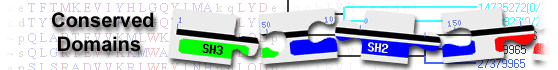
This image shows a graphical summary of conserved domains identified on the query sequence.
The Show Concise/Full Display button at the top of the page can be used to select the desired level of detail: only top scoring hits
(labeled illustration) or all hits
(labeled illustration).
Domains are color coded according to superfamilies
to which they have been assigned. Hits with scores that pass a domain-specific threshold
(specific hits) are drawn in bright colors.
Others (non-specific hits) and
superfamily placeholders are drawn in pastel colors.
if a domain or superfamily has been annotated with functional sites (conserved features),
they are mapped to the query sequence and indicated through sets of triangles
with the same color and shade of the domain or superfamily that provides the annotation. Mouse over the colored bars or triangles to see descriptions of the domains and features.
click on the bars or triangles to view your query sequence embedded in a multiple sequence alignment of the proteins used to develop the corresponding domain model.
The table lists conserved domains identified on the query sequence. Click on the plus sign (+) on the left to display full descriptions, alignments, and scores.
Click on the domain model's accession number to view the multiple sequence alignment of the proteins used to develop the corresponding domain model.
To view your query sequence embedded in that multiple sequence alignment, click on the colored bars in the Graphical Summary portion of the search results page,
or click on the triangles, if present, that represent functional sites (conserved features)
mapped to the query sequence.
Concise Display shows only the best scoring domain model, in each hit category listed below except non-specific hits, for each region on the query sequence.
(labeled illustration) Standard Display shows only the best scoring domain model from each source, in each hit category listed below for each region on the query sequence.
(labeled illustration) Full Display shows all domain models, in each hit category below, that meet or exceed the RPS-BLAST threshold for statistical significance.
(labeled illustration) Four types of hits can be shown, as available,
for each region on the query sequence:
specific hits meet or exceed a domain-specific e-value threshold
(illustrated example)
and represent a very high confidence that the query sequence belongs to the same protein family as the sequences use to create the domain model
non-specific hits
meet or exceed the RPS-BLAST threshold for statistical significance (default E-value cutoff of 0.01, or an E-value selected by user via the
advanced search options)
the domain superfamily to which the specific and non-specific hits belong
multi-domain models that were computationally detected and are likely to contain multiple single domains
Retrieve proteins that contain one or more of the domains present in the query sequence, using the Conserved Domain Architecture Retrieval Tool
(CDART).
Modify your query to search against a different database and/or use advanced search options