trans-2-enoyl-CoA reductase (TER) family protein such as enoyl-[acyl-carrier-protein] reductase FabV and trans-2-enoyl-CoA reductase, which are both involved in fatty acid synthesis
Trans-2-enoyl-CoA reductase catalytic region; This family of trans-2-enoyl-CoA reductases, EC: ...
82-317
5.10e-162
Trans-2-enoyl-CoA reductase catalytic region; This family of trans-2-enoyl-CoA reductases, EC:1.3.1.44, carries the the catalytic sites of the enzyme, characterized by the conserved sequence motifs: YNThhhFxK, and YShAPxR. In Euglena where the enzyme has been characterized it catalyzes the reduction of enoyl-CoA to acyl-CoA in an unusual fatty acid pathway in mitochondria. the whole path performs a malonyl-CoA independent synthesis of fatty acids leading to accumulation of wax esters, which serve as the sink for electrons stemming from glycolytic ATP synthesis and pyruvate oxidation.
Pssm-ID: 463504 Cd Length: 236 Bit Score: 454.26 E-value: 5.10e-162
mannitol dehydrogenase (MDH)-like, classical (c) SDRs; NADP-mannitol dehydrogenase catalyzes the conversion of fructose to mannitol, an acyclic 6-carbon sugar. MDH is a tetrameric member of the SDR family. This subgroup also includes various other tetrameric SDRs, including Pichia stipitis D-arabinitol dehydrogenase (aka polyol dehydrogenase), Candida albicans Sou1p, a sorbose reductase, and Candida parapsilosis (S)-specific carbonyl reductase (SCR, aka S-specific alcohol dehydrogenase) which catalyzes the enantioselective reduction of 2-hydroxyacetophenone into (S)-1-phenyl-1,2-ethanediol. SDRs are a functionally diverse family of oxidoreductases that have a single domain with a structurally conserved Rossmann fold (alpha/beta folding pattern with a central beta-sheet), an NAD(P)(H)-binding region, and a structurally diverse C-terminal region. Classical SDRs are typically about 250 residues long, while extended SDRS are approximately 350 residues. Sequence identity between different SDR enzymes are typically in the 15-30% range, but the enzymes share the Rossmann fold NAD-binding motif and characteristic NAD-binding and catalytic sequence patterns. These enzymes have a 3-glycine N-terminal NAD(P)(H)-binding pattern (typically, TGxxxGxG in classical SDRs and TGxxGxxG in extended SDRs), while substrate binding is in the C-terminal region. A critical catalytic Tyr residue (Tyr-151, human 15-hydroxyprostaglandin dehydrogenase (15-PGDH) numbering), is often found in a conserved YXXXK pattern. In addition to the Tyr and Lys, there is often an upstream Ser (Ser-138, 15-PGDH numbering) and/or an Asn (Asn-107, 15-PGDH numbering) or additional Ser, contributing to the active site. Substrates for these enzymes include sugars, steroids, alcohols, and aromatic compounds. The standard reaction mechanism is a proton relay involving the conserved Tyr and Lys, as well as Asn (or Ser).
Pssm-ID: 187610 [Multi-domain] Cd Length: 252 Bit Score: 38.08 E-value: 5.69e-03
Trans-2-enoyl-CoA reductase catalytic region; This family of trans-2-enoyl-CoA reductases, EC: ...
82-317
5.10e-162
Trans-2-enoyl-CoA reductase catalytic region; This family of trans-2-enoyl-CoA reductases, EC:1.3.1.44, carries the the catalytic sites of the enzyme, characterized by the conserved sequence motifs: YNThhhFxK, and YShAPxR. In Euglena where the enzyme has been characterized it catalyzes the reduction of enoyl-CoA to acyl-CoA in an unusual fatty acid pathway in mitochondria. the whole path performs a malonyl-CoA independent synthesis of fatty acids leading to accumulation of wax esters, which serve as the sink for electrons stemming from glycolytic ATP synthesis and pyruvate oxidation.
Pssm-ID: 463504 Cd Length: 236 Bit Score: 454.26 E-value: 5.10e-162
NAD(P)H binding domain of trans-2-enoyl-CoA reductase; This family carries the region of the ...
2-80
1.85e-38
NAD(P)H binding domain of trans-2-enoyl-CoA reductase; This family carries the region of the enzyme trans-2-enoyl-CoA reductase, EC:1.3.1.44, which binds NAD(P)H. The activity of the enzyme was characterized in Euglena where an unusual fatty acid synthesis path-way in the mitochondria performs a malonyl-CoA independent synthesis of fatty acids leading to accumulation of wax esters, which serve as the sink for electrons stemming from glycolytic ATP synthesis and pyruvate oxidation. The full enzyme catalyzes the reduction of enoyl-CoA to acyl-CoA. The binding site is conserved as GA/CSpGYG, where p is any polar residue.
Pssm-ID: 432420 [Multi-domain] Cd Length: 78 Bit Score: 132.96 E-value: 1.85e-38
Enoyl reductase FAD binding domain; This family carries the region of the enzyme ...
326-389
1.51e-35
Enoyl reductase FAD binding domain; This family carries the region of the enzyme trans-2-enoyl-CoA reductase, at the very C-terminus, that binds to FAD. The activity was characterized in Euglena where an unusual fatty acid synthesis path-way in mitochondria performs a malonyl-CoA independent synthesis of fatty acids leading to accumulation of wax esters, which serve as the sink for electrons stemming from glycolytic ATP synthesis and pyruvate oxidation. The full enzyme catalyzes the reduction of enoyl-CoA to acyl-CoA. The conserved region is seen as the motif FGFxxxxxDY.
Pssm-ID: 462075 [Multi-domain] Cd Length: 64 Bit Score: 124.88 E-value: 1.51e-35
mannitol dehydrogenase (MDH)-like, classical (c) SDRs; NADP-mannitol dehydrogenase catalyzes the conversion of fructose to mannitol, an acyclic 6-carbon sugar. MDH is a tetrameric member of the SDR family. This subgroup also includes various other tetrameric SDRs, including Pichia stipitis D-arabinitol dehydrogenase (aka polyol dehydrogenase), Candida albicans Sou1p, a sorbose reductase, and Candida parapsilosis (S)-specific carbonyl reductase (SCR, aka S-specific alcohol dehydrogenase) which catalyzes the enantioselective reduction of 2-hydroxyacetophenone into (S)-1-phenyl-1,2-ethanediol. SDRs are a functionally diverse family of oxidoreductases that have a single domain with a structurally conserved Rossmann fold (alpha/beta folding pattern with a central beta-sheet), an NAD(P)(H)-binding region, and a structurally diverse C-terminal region. Classical SDRs are typically about 250 residues long, while extended SDRS are approximately 350 residues. Sequence identity between different SDR enzymes are typically in the 15-30% range, but the enzymes share the Rossmann fold NAD-binding motif and characteristic NAD-binding and catalytic sequence patterns. These enzymes have a 3-glycine N-terminal NAD(P)(H)-binding pattern (typically, TGxxxGxG in classical SDRs and TGxxGxxG in extended SDRs), while substrate binding is in the C-terminal region. A critical catalytic Tyr residue (Tyr-151, human 15-hydroxyprostaglandin dehydrogenase (15-PGDH) numbering), is often found in a conserved YXXXK pattern. In addition to the Tyr and Lys, there is often an upstream Ser (Ser-138, 15-PGDH numbering) and/or an Asn (Asn-107, 15-PGDH numbering) or additional Ser, contributing to the active site. Substrates for these enzymes include sugars, steroids, alcohols, and aromatic compounds. The standard reaction mechanism is a proton relay involving the conserved Tyr and Lys, as well as Asn (or Ser).
Pssm-ID: 187610 [Multi-domain] Cd Length: 252 Bit Score: 38.08 E-value: 5.69e-03
Database: CDSEARCH/cdd Low complexity filter: no Composition Based Adjustment: yes E-value threshold: 0.01
References:
Wang J et al. (2023), "The conserved domain database in 2023", Nucleic Acids Res.51(D)384-8.
Lu S et al. (2020), "The conserved domain database in 2020", Nucleic Acids Res.48(D)265-8.
Marchler-Bauer A et al. (2017), "CDD/SPARCLE: functional classification of proteins via subfamily domain architectures.", Nucleic Acids Res.45(D)200-3.
of the residues that compose this conserved feature have been mapped to the query sequence.
Click on the triangle to view details about the feature, including a multiple sequence alignment
of your query sequence and the protein sequences used to curate the domain model,
where hash marks (#) above the aligned sequences show the location of the conserved feature residues.
The thumbnail image, if present, provides an approximate view of the feature's location in 3 dimensions.
Click on the triangle for interactive 3D structure viewing options.
Functional characterization of the conserved domain architecture found on the query.
Click here to see more details.
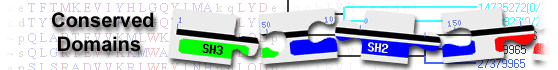
This image shows a graphical summary of conserved domains identified on the query sequence.
The Show Concise/Full Display button at the top of the page can be used to select the desired level of detail: only top scoring hits
(labeled illustration) or all hits
(labeled illustration).
Domains are color coded according to superfamilies
to which they have been assigned. Hits with scores that pass a domain-specific threshold
(specific hits) are drawn in bright colors.
Others (non-specific hits) and
superfamily placeholders are drawn in pastel colors.
if a domain or superfamily has been annotated with functional sites (conserved features),
they are mapped to the query sequence and indicated through sets of triangles
with the same color and shade of the domain or superfamily that provides the annotation. Mouse over the colored bars or triangles to see descriptions of the domains and features.
click on the bars or triangles to view your query sequence embedded in a multiple sequence alignment of the proteins used to develop the corresponding domain model.
The table lists conserved domains identified on the query sequence. Click on the plus sign (+) on the left to display full descriptions, alignments, and scores.
Click on the domain model's accession number to view the multiple sequence alignment of the proteins used to develop the corresponding domain model.
To view your query sequence embedded in that multiple sequence alignment, click on the colored bars in the Graphical Summary portion of the search results page,
or click on the triangles, if present, that represent functional sites (conserved features)
mapped to the query sequence.
Concise Display shows only the best scoring domain model, in each hit category listed below except non-specific hits, for each region on the query sequence.
(labeled illustration) Standard Display shows only the best scoring domain model from each source, in each hit category listed below for each region on the query sequence.
(labeled illustration) Full Display shows all domain models, in each hit category below, that meet or exceed the RPS-BLAST threshold for statistical significance.
(labeled illustration) Four types of hits can be shown, as available,
for each region on the query sequence:
specific hits meet or exceed a domain-specific e-value threshold
(illustrated example)
and represent a very high confidence that the query sequence belongs to the same protein family as the sequences use to create the domain model
non-specific hits
meet or exceed the RPS-BLAST threshold for statistical significance (default E-value cutoff of 0.01, or an E-value selected by user via the
advanced search options)
the domain superfamily to which the specific and non-specific hits belong
multi-domain models that were computationally detected and are likely to contain multiple single domains
Retrieve proteins that contain one or more of the domains present in the query sequence, using the Conserved Domain Architecture Retrieval Tool
(CDART).
Modify your query to search against a different database and/or use advanced search options