2,3,4,5-tetrahydropyridine-2,6-dicarboxylate N-succinyltransferase catalyzes the conversion of the cyclic tetrahydrodipicolinate (THDP) into the acyclic N-succinyl-L-2-amino-6-oxopimelate using succinyl-CoA
2,3,4,5-tetrahydropyridine-2,6-dicarboxylate N-succinyltransferase; 2,3,4,5-tetrahydropyridine-2,6-dicarboxylate N-succinyltransferase (DapD) is involved in the succinylated branch of the "lysine biosynthesis via diaminopimelate (DAP)" pathway (GenProp0125). This model represents a clade of DapD sequences most closely related to the actinobacterial DapD family represented by the TIGR03535 model. All of the genes evaluated for the seed of this model are found in genomes where the downstream desuccinylase is present, but known DapD genes are absent. Additionally, many of the genes identified by this model are found proximal to genes involved in this lysine biosynthesis pathway.
:
Pssm-ID: 211834 [Multi-domain] Cd Length: 341 Bit Score: 636.46 E-value: 0e+00
2,3,4,5-tetrahydropyridine-2,6-dicarboxylate N-succinyltransferase; 2,3,4,5-tetrahydropyridine-2,6-dicarboxylate N-succinyltransferase (DapD) is involved in the succinylated branch of the "lysine biosynthesis via diaminopimelate (DAP)" pathway (GenProp0125). This model represents a clade of DapD sequences most closely related to the actinobacterial DapD family represented by the TIGR03535 model. All of the genes evaluated for the seed of this model are found in genomes where the downstream desuccinylase is present, but known DapD genes are absent. Additionally, many of the genes identified by this model are found proximal to genes involved in this lysine biosynthesis pathway.
Pssm-ID: 211834 [Multi-domain] Cd Length: 341 Bit Score: 636.46 E-value: 0e+00
Putative 2,3,4,5-tetrahydropyridine-2,6-dicarboxylate (THDP) N-succinyltransferase (THP succinyltransferase), C-terminal left-handed parallel alpha-helix (LbH) domain: This group is composed of mostly uncharacterized proteins containing an N-terminal domain of unknown function and a C-terminal LbH domain with similarity to THP succinyltransferase LbH. THP succinyltransferase catalyzes the conversion of tetrahydrodipicolinate and succinyl-CoA to N-succinyltetrahydrodipicolinate and CoA. It is the committed step in the succinylase pathway by which bacteria synthesize L-lysine and meso-diaminopimelate, a component of peptidoglycan. The enzyme is trimeric and displays the left-handed parallel alpha-helix (LbH) structural motif encoded by the hexapeptide repeat motif.
Pssm-ID: 100054 Cd Length: 147 Bit Score: 241.55 E-value: 1.71e-80
Tetrahydrodipicolinate N-succinyltransferase [Amino acid transport and metabolism]; ...
129-340
7.28e-65
Tetrahydrodipicolinate N-succinyltransferase [Amino acid transport and metabolism]; Tetrahydrodipicolinate N-succinyltransferase is part of the Pathway/BioSystem: Lysine biosynthesis
Pssm-ID: 441774 [Multi-domain] Cd Length: 268 Bit Score: 206.12 E-value: 7.28e-65
2,3,4,5-tetrahydropyridine-2,6-dicarboxylate N-succinyltransferase; 2,3,4,5-tetrahydropyridine-2,6-dicarboxylate N-succinyltransferase (DapD) is involved in the succinylated branch of the "lysine biosynthesis via diaminopimelate (DAP)" pathway (GenProp0125). This model represents a clade of DapD sequences most closely related to the actinobacterial DapD family represented by the TIGR03535 model. All of the genes evaluated for the seed of this model are found in genomes where the downstream desuccinylase is present, but known DapD genes are absent. Additionally, many of the genes identified by this model are found proximal to genes involved in this lysine biosynthesis pathway.
Pssm-ID: 211834 [Multi-domain] Cd Length: 341 Bit Score: 636.46 E-value: 0e+00
2,3,4,5-tetrahydropyridine-2,6-dicarboxylate N-succinyltransferase; This enzyme is part of the ...
3-340
1.57e-134
2,3,4,5-tetrahydropyridine-2,6-dicarboxylate N-succinyltransferase; This enzyme is part of the diaminopimelate pathway of lysine biosynthesis. This model represents a clade of the enzyme specific to Actinobacteria. Alternate name: tetrahydrodipicolinate N-succinyltransferase.
Pssm-ID: 274635 [Multi-domain] Cd Length: 319 Bit Score: 385.64 E-value: 1.57e-134
Putative 2,3,4,5-tetrahydropyridine-2,6-dicarboxylate (THDP) N-succinyltransferase (THP succinyltransferase), C-terminal left-handed parallel alpha-helix (LbH) domain: This group is composed of mostly uncharacterized proteins containing an N-terminal domain of unknown function and a C-terminal LbH domain with similarity to THP succinyltransferase LbH. THP succinyltransferase catalyzes the conversion of tetrahydrodipicolinate and succinyl-CoA to N-succinyltetrahydrodipicolinate and CoA. It is the committed step in the succinylase pathway by which bacteria synthesize L-lysine and meso-diaminopimelate, a component of peptidoglycan. The enzyme is trimeric and displays the left-handed parallel alpha-helix (LbH) structural motif encoded by the hexapeptide repeat motif.
Pssm-ID: 100054 Cd Length: 147 Bit Score: 241.55 E-value: 1.71e-80
Tetrahydrodipicolinate N-succinyltransferase [Amino acid transport and metabolism]; ...
129-340
7.28e-65
Tetrahydrodipicolinate N-succinyltransferase [Amino acid transport and metabolism]; Tetrahydrodipicolinate N-succinyltransferase is part of the Pathway/BioSystem: Lysine biosynthesis
Pssm-ID: 441774 [Multi-domain] Cd Length: 268 Bit Score: 206.12 E-value: 7.28e-65
Left-handed parallel beta-Helix (LbetaH or LbH) domain: The alignment contains 5 turns, each ...
209-282
4.73e-08
Left-handed parallel beta-Helix (LbetaH or LbH) domain: The alignment contains 5 turns, each containing three imperfect tandem repeats of a hexapeptide repeat motif (X-[STAV]-X-[LIV]-[GAED]-X). Proteins containing hexapeptide repeats are often enzymes showing acyltransferase activity, however, some subfamilies in this hierarchy also show activities related to ion transport or translation initiation. Many are trimeric in their active forms.
Pssm-ID: 100038 [Multi-domain] Cd Length: 78 Bit Score: 49.55 E-value: 4.73e-08
2,3,4,5-tetrahydropyridine-2,6-dicarboxylate (THDP) N-succinyltransferase (also called THP ...
178-287
7.77e-08
2,3,4,5-tetrahydropyridine-2,6-dicarboxylate (THDP) N-succinyltransferase (also called THP succinyltransferase): THDP N-succinyltransferase catalyzes the conversion of tetrahydrodipicolinate and succinyl-CoA to N-succinyltetrahydrodipicolinate and CoA. It is the committed step in the succinylase pathway by which bacteria synthesize L-lysine and meso-diaminopimelate, a component of peptidoglycan. The enzyme is homotrimeric and each subunit contains an N-terminal region with alpha helices and hairpin loops, as well as a C-terminal region with a left-handed parallel alpha-helix (LbH) structural motif encoded by hexapeptide repeat motifs.
Pssm-ID: 100041 [Multi-domain] Cd Length: 139 Bit Score: 50.84 E-value: 7.77e-08
Putative Acyltransferase (AT), Left-handed parallel beta-Helix (LbH) domain; This group is ...
181-288
1.31e-04
Putative Acyltransferase (AT), Left-handed parallel beta-Helix (LbH) domain; This group is composed of mostly uncharacterized proteins containing an N-terminal helical subdomain followed by a LbH domain. The alignment contains 6 turns, each containing three imperfect tandem repeats of a hexapeptide repeat motif (X-[STAV]-X-[LIV]-[GAED]-X). Proteins containing hexapeptide repeats are often enzymes showing acyltransferase activity. A few members are identified as NeuD, a sialic acid (Sia) O-acetyltransferase that is required for Sia synthesis and surface polysaccharide sialylation.
Pssm-ID: 100050 [Multi-domain] Cd Length: 197 Bit Score: 42.47 E-value: 1.31e-04
Tetrahydrodipicolinate N-succinyltransferase N-terminal; This is the N-terminal domain of 2,3, ...
2-126
3.23e-04
Tetrahydrodipicolinate N-succinyltransferase N-terminal; This is the N-terminal domain of 2,3,4,5-tetrahydropyridine-2,6-dicarboxylate N-succinyltransferase.
Pssm-ID: 339376 Cd Length: 167 Bit Score: 40.72 E-value: 3.23e-04
Serine acetyltransferase (SAT): SAT catalyzes the CoA-dependent acetylation of the side chain ...
189-288
5.20e-03
Serine acetyltransferase (SAT): SAT catalyzes the CoA-dependent acetylation of the side chain hydroxyl group of L-serine to form O-acetylserine, as the first step of a two-step biosynthetic pathway in bacteria and plants leading to the formation of L-cysteine. This reaction represents a key metabolic point of regulation for the cysteine biosynthetic pathway due to its feedback inhibition by cysteine. The enzyme is a 175 kDa homohexamer, composed of a dimer of homotrimers. Each subunit contains an N-terminal alpha helical region and a C-terminal left-handed beta-helix (LbH) subdomain with 5 turns, each containing a hexapeptide repeat motif characteristic of the acyltransferase superfamily of enzymes. The trimer interface mainly involves the C-terminal LbH subdomain while the dimer (of trimers) interface is mediated by the N-terminal alpha helical subdomain.
Pssm-ID: 100045 [Multi-domain] Cd Length: 101 Bit Score: 35.88 E-value: 5.20e-03
Database: CDSEARCH/cdd Low complexity filter: no Composition Based Adjustment: yes E-value threshold: 0.01
References:
Wang J et al. (2023), "The conserved domain database in 2023", Nucleic Acids Res.51(D)384-8.
Lu S et al. (2020), "The conserved domain database in 2020", Nucleic Acids Res.48(D)265-8.
Marchler-Bauer A et al. (2017), "CDD/SPARCLE: functional classification of proteins via subfamily domain architectures.", Nucleic Acids Res.45(D)200-3.
of the residues that compose this conserved feature have been mapped to the query sequence.
Click on the triangle to view details about the feature, including a multiple sequence alignment
of your query sequence and the protein sequences used to curate the domain model,
where hash marks (#) above the aligned sequences show the location of the conserved feature residues.
The thumbnail image, if present, provides an approximate view of the feature's location in 3 dimensions.
Click on the triangle for interactive 3D structure viewing options.
Functional characterization of the conserved domain architecture found on the query.
Click here to see more details.
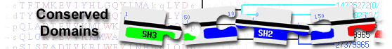
This image shows a graphical summary of conserved domains identified on the query sequence.
The Show Concise/Full Display button at the top of the page can be used to select the desired level of detail: only top scoring hits
(labeled illustration) or all hits
(labeled illustration).
Domains are color coded according to superfamilies
to which they have been assigned. Hits with scores that pass a domain-specific threshold
(specific hits) are drawn in bright colors.
Others (non-specific hits) and
superfamily placeholders are drawn in pastel colors.
if a domain or superfamily has been annotated with functional sites (conserved features),
they are mapped to the query sequence and indicated through sets of triangles
with the same color and shade of the domain or superfamily that provides the annotation. Mouse over the colored bars or triangles to see descriptions of the domains and features.
click on the bars or triangles to view your query sequence embedded in a multiple sequence alignment of the proteins used to develop the corresponding domain model.
The table lists conserved domains identified on the query sequence. Click on the plus sign (+) on the left to display full descriptions, alignments, and scores.
Click on the domain model's accession number to view the multiple sequence alignment of the proteins used to develop the corresponding domain model.
To view your query sequence embedded in that multiple sequence alignment, click on the colored bars in the Graphical Summary portion of the search results page,
or click on the triangles, if present, that represent functional sites (conserved features)
mapped to the query sequence.
Concise Display shows only the best scoring domain model, in each hit category listed below except non-specific hits, for each region on the query sequence.
(labeled illustration) Standard Display shows only the best scoring domain model from each source, in each hit category listed below for each region on the query sequence.
(labeled illustration) Full Display shows all domain models, in each hit category below, that meet or exceed the RPS-BLAST threshold for statistical significance.
(labeled illustration) Four types of hits can be shown, as available,
for each region on the query sequence:
specific hits meet or exceed a domain-specific e-value threshold
(illustrated example)
and represent a very high confidence that the query sequence belongs to the same protein family as the sequences use to create the domain model
non-specific hits
meet or exceed the RPS-BLAST threshold for statistical significance (default E-value cutoff of 0.01, or an E-value selected by user via the
advanced search options)
the domain superfamily to which the specific and non-specific hits belong
multi-domain models that were computationally detected and are likely to contain multiple single domains
Retrieve proteins that contain one or more of the domains present in the query sequence, using the Conserved Domain Architecture Retrieval Tool
(CDART).
Modify your query to search against a different database and/or use advanced search options