

Cna B-type domain-containing protein, partial [Bacillus toyonensis]
Protein Classification

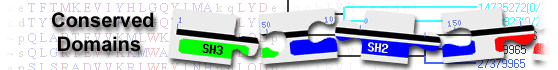
List of domain hits
| Name | Accession | Description | Interval | E-value | |||
| Cna_B | pfam05738 | Cna protein B-type domain; This domain is found in Staphylococcus aureus collagen-binding ... |
1-51 | 1.49e-19 | |||
Cna protein B-type domain; This domain is found in Staphylococcus aureus collagen-binding surface protein. The structure of the repetitive B-region has been solved and forms a beta sandwich structure. : Pssm-ID: 461726 [Multi-domain] Cd Length: 88 Bit Score: 77.29 E-value: 1.49e-19
|
|||||||
| SCO1860_LAETG super family | cl49495 | SCO1860 family LAETG-anchored protein; Members of this poorly characterized family, including ... |
41-148 | 2.29e-06 | |||
SCO1860 family LAETG-anchored protein; Members of this poorly characterized family, including SCO1860 from Streptomyces coelicolor, are surface proteins whose C-terminus contains a variant type sortase recognition and cleavage sorting signal. The sorting signal motif, LAETG, is compatible with processing by a SrtE family sortase enzyme. The actual alignment was detected with superfamily member NF041527: Pssm-ID: 469412 [Multi-domain] Cd Length: 305 Bit Score: 45.50 E-value: 2.29e-06
|
|||||||
| Name | Accession | Description | Interval | E-value | |||
| Cna_B | pfam05738 | Cna protein B-type domain; This domain is found in Staphylococcus aureus collagen-binding ... |
1-51 | 1.49e-19 | |||
Cna protein B-type domain; This domain is found in Staphylococcus aureus collagen-binding surface protein. The structure of the repetitive B-region has been solved and forms a beta sandwich structure. Pssm-ID: 461726 [Multi-domain] Cd Length: 88 Bit Score: 77.29 E-value: 1.49e-19
|
|||||||
| CollagenBindB | cd00222 | Repeat unit of collagen-binding protein domain B; The collagen-binding protein mediates ... |
1-52 | 1.92e-16 | |||
Repeat unit of collagen-binding protein domain B; The collagen-binding protein mediates bacterial adherence to collagen; the primary sequence has a non-repetitive, collagen-binding A region, followed by instances of this B region repetitive unit. The B region has one to four 23 kDa repeat units (B1-B4), which have been suggested to serve as 'stalks' that project the A region from the bacterial surface and thus facilitate bacterial adherence to collagen. Each B repeat unit has two highly similar domains (D1 and D2) placed side-by-side; both D1 and D2 are included in this model. They exhibit a unique inverse IgG-like domain fold. Pssm-ID: 212461 [Multi-domain] Cd Length: 92 Bit Score: 69.63 E-value: 1.92e-16
|
|||||||
| SCO1860_LAETG | NF041527 | SCO1860 family LAETG-anchored protein; Members of this poorly characterized family, including ... |
41-148 | 2.29e-06 | |||
SCO1860 family LAETG-anchored protein; Members of this poorly characterized family, including SCO1860 from Streptomyces coelicolor, are surface proteins whose C-terminus contains a variant type sortase recognition and cleavage sorting signal. The sorting signal motif, LAETG, is compatible with processing by a SrtE family sortase enzyme. Pssm-ID: 469412 [Multi-domain] Cd Length: 305 Bit Score: 45.50 E-value: 2.29e-06
|
|||||||
| LPXTG_anchor | TIGR01167 | LPXTG-motif cell wall anchor domain; This model describes the LPXTG motif-containing region ... |
115-148 | 2.33e-05 | |||
LPXTG-motif cell wall anchor domain; This model describes the LPXTG motif-containing region found at the C-terminus of many surface proteins of Streptococcus and Streptomyces species. Cleavage between the Thr and Gly by sortase or a related enzyme leads to covalent anchoring at the new C-terminal Thr to the cell wall. Hits that do not lie at the C-terminus or are not found in Gram-positive bacteria are probably false-positive. A common feature of this proteins containing this domain appears to be a high proportion of charged and zwitterionic residues immediatedly upstream of the LPXTG motif. This model differs from other descriptions of the LPXTG region by including a portion of that upstream charged region. [Cell envelope, Other] Pssm-ID: 273478 [Multi-domain] Cd Length: 34 Bit Score: 39.38 E-value: 2.33e-05
|
|||||||
| PRK09418 | PRK09418 | bifunctional 2',3'-cyclic-nucleotide 2'-phosphodiesterase/3'-nucleotidase; |
50-146 | 7.48e-05 | |||
bifunctional 2',3'-cyclic-nucleotide 2'-phosphodiesterase/3'-nucleotidase; Pssm-ID: 236504 [Multi-domain] Cd Length: 780 Bit Score: 41.62 E-value: 7.48e-05
|
|||||||
| Gram_pos_anchor | pfam00746 | LPXTG cell wall anchor motif; |
106-146 | 5.61e-03 | |||
LPXTG cell wall anchor motif; Pssm-ID: 366278 [Multi-domain] Cd Length: 43 Bit Score: 32.90 E-value: 5.61e-03
|
|||||||
| PRK14950 | PRK14950 | DNA polymerase III subunits gamma and tau; Provisional |
51-107 | 8.48e-03 | |||
DNA polymerase III subunits gamma and tau; Provisional Pssm-ID: 237864 [Multi-domain] Cd Length: 585 Bit Score: 35.56 E-value: 8.48e-03
|
|||||||
| Blast search parameters | ||||
|
