

hepatitis A virus cellular receptor 2 isoform X1 [Gallus gallus]
Protein Classification

immunoglobulin domain-containing family protein( domain architecture ID 34076)
immunoglobulin (Ig) domain-containing family protein is a member of a large superfamily containing cell surface antigen receptors, co-receptors and co-stimulatory molecules of the immune system, molecules involved in antigen presentation to lymphocytes, cell adhesion molecules, certain cytokine receptors and intracellular muscle proteins; immunoglobulin domains are typically divided into 4 main classes based on their structures and sequences: the Variable (V), Constant 1 (C1), Constant 2 (C2), and Intermediate (I) sets
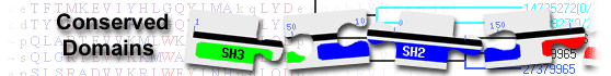
List of domain hits
| Name | Accession | Description | Interval | E-value | |||
| Ig super family | cl11960 | Immunoglobulin domain; The members here are composed of the immunoglobulin (Ig) domain found ... |
25-129 | 1.35e-44 | |||
Immunoglobulin domain; The members here are composed of the immunoglobulin (Ig) domain found in the Ig superfamily. The Ig superfamily is a heterogenous group of proteins, built on a common fold comprised of a sandwich of two beta sheets. Members of this group are components of immunoglobulin, neuroglia, cell surface glycoproteins, including T-cell receptors, CD2, CD4, CD8, and membrane glycoproteins, including butyrophilin and chondroitin sulfate proteoglycan core protein. A predominant feature of most Ig domains is a disulfide bridge connecting the two beta-sheets with a tryptophan residue packed against the disulfide bond. Ig superfamily (IgSF) domains can be divided into 4 main classes based on their structures and sequences: the Variable (V), Constant 1 (C1), Constant 2 (C2), and Intermediate (I) sets. Typically, the V-set domains have A, B, E, and D strands in one sheet and A', G, F, C, C' and C" in the other. The structures in C1-set are smaller than those in the V-set; they have one beta sheet that is formed by strands A, B, E, and D and the other by strands G, F, C, and C'. Moreover, a C1-set Ig domain contains a short C' strand (three residues) and lacks A' and C" strand. Unlike other Ig domain sets, C2-set structures do not have a D strand. Like the V-set Ig domains, members of the I-set have a discontinuous A strand, but lack a C" strand. The actual alignment was detected with superfamily member cd20982: Pssm-ID: 472250 Cd Length: 107 Bit Score: 147.22 E-value: 1.35e-44
|
|||||||
| Name | Accession | Description | Interval | E-value | |||
| IgV_TIM-3_like | cd20982 | Immunoglobulin Variable (IgV) domain of T cell Immunoglobulin Domain and Mucin Domain 3 (Tim-3) ... |
25-129 | 1.35e-44 | |||
Immunoglobulin Variable (IgV) domain of T cell Immunoglobulin Domain and Mucin Domain 3 (Tim-3), and similar domains; The members here are composed of the immunoglobulin variable (IgV) domain of T cell immunoglobulin domain and mucin domain 3 (Tim-3; also known as Hepatitis A virus cellular receptor 2 (HAVcr-2) and Cluster of Differentiation 366 (CD366)) and similar proteins. TIM-3 is a checkpoint inhibitor in immune responses to tumors, as well as involved in chronic viral infections. Thus, Tim-3 has emerged as one of most promising immune checkpoint targets for cancer immunotherapy. Tim-3 is highly expressed on Th1 lymphocytes and CD11b(+) macrophages and is upregulated on activated T and myeloid cells. TIM-3 regulates macrophage, activation and inhibits Th1 mediated immune responses to promote immunological tolerance. There are three TIM family members in humans (TIM-1, TIM-3, and TIM-4) and eight members in mice (TIM-1 to TIM-8). The IgV domain of human TIM-3 has been shown to bind ligands such as carcinoembryonic antigen cell adhesion molecule 1 (CEACAM1), high mobility group protein B1 (HMGB1)and galectin-9 (GAL9). The binding of GAL9 to TIM-3 can negatively regulate Th1 immune response, enhance immune tolerance and inhibit anti#tumor immunity. Dysregulation of the TIM-3/GAL9 pathway is implicated in numerous chronic autoimmune diseases, such as multiple sclerosis and systemic lupus erythematosus. Pssm-ID: 409574 Cd Length: 107 Bit Score: 147.22 E-value: 1.35e-44
|
|||||||
| V-set | pfam07686 | Immunoglobulin V-set domain; This domain is found in antibodies as well as neural protein P0 ... |
25-128 | 1.02e-10 | |||
Immunoglobulin V-set domain; This domain is found in antibodies as well as neural protein P0 and CTL4 amongst others. Pssm-ID: 462230 Cd Length: 109 Bit Score: 57.85 E-value: 1.02e-10
|
|||||||
| IG_like | smart00410 | Immunoglobulin like; IG domains that cannot be classified into one of IGv1, IGc1, IGc2, IG. |
25-127 | 1.47e-06 | |||
Immunoglobulin like; IG domains that cannot be classified into one of IGv1, IGc1, IGc2, IG. Pssm-ID: 214653 [Multi-domain] Cd Length: 85 Bit Score: 45.57 E-value: 1.47e-06
|
|||||||
| Name | Accession | Description | Interval | E-value | |||
| IgV_TIM-3_like | cd20982 | Immunoglobulin Variable (IgV) domain of T cell Immunoglobulin Domain and Mucin Domain 3 (Tim-3) ... |
25-129 | 1.35e-44 | |||
Immunoglobulin Variable (IgV) domain of T cell Immunoglobulin Domain and Mucin Domain 3 (Tim-3), and similar domains; The members here are composed of the immunoglobulin variable (IgV) domain of T cell immunoglobulin domain and mucin domain 3 (Tim-3; also known as Hepatitis A virus cellular receptor 2 (HAVcr-2) and Cluster of Differentiation 366 (CD366)) and similar proteins. TIM-3 is a checkpoint inhibitor in immune responses to tumors, as well as involved in chronic viral infections. Thus, Tim-3 has emerged as one of most promising immune checkpoint targets for cancer immunotherapy. Tim-3 is highly expressed on Th1 lymphocytes and CD11b(+) macrophages and is upregulated on activated T and myeloid cells. TIM-3 regulates macrophage, activation and inhibits Th1 mediated immune responses to promote immunological tolerance. There are three TIM family members in humans (TIM-1, TIM-3, and TIM-4) and eight members in mice (TIM-1 to TIM-8). The IgV domain of human TIM-3 has been shown to bind ligands such as carcinoembryonic antigen cell adhesion molecule 1 (CEACAM1), high mobility group protein B1 (HMGB1)and galectin-9 (GAL9). The binding of GAL9 to TIM-3 can negatively regulate Th1 immune response, enhance immune tolerance and inhibit anti#tumor immunity. Dysregulation of the TIM-3/GAL9 pathway is implicated in numerous chronic autoimmune diseases, such as multiple sclerosis and systemic lupus erythematosus. Pssm-ID: 409574 Cd Length: 107 Bit Score: 147.22 E-value: 1.35e-44
|
|||||||
| V-set | pfam07686 | Immunoglobulin V-set domain; This domain is found in antibodies as well as neural protein P0 ... |
25-128 | 1.02e-10 | |||
Immunoglobulin V-set domain; This domain is found in antibodies as well as neural protein P0 and CTL4 amongst others. Pssm-ID: 462230 Cd Length: 109 Bit Score: 57.85 E-value: 1.02e-10
|
|||||||
| IgV_pIgR_like | cd05716 | Immunoglobulin (Ig)-like domain in the polymeric Ig receptor (pIgR) and similar proteins; The ... |
15-128 | 2.76e-09 | |||
Immunoglobulin (Ig)-like domain in the polymeric Ig receptor (pIgR) and similar proteins; The members here are composed of the immunoglobulin (Ig)-like domain in the polymeric Ig receptor (pIgR) and similar proteins. pIgR delivers dimeric IgA and pentameric IgM to mucosal secretions. Polymeric immunoglobulin (pIgs) are the first defense against pathogens and toxins. IgA and IgM can form polymers via an 18-residue extension at their C-termini referred to as the tailpiece. pIgR transports pIgs across mucosal epithelia into mucosal secretions. Human pIgR is a glycosylated type I transmembrane protein, comprised of a 620-residue extracellular region, a 23-residue transmembrane region, and a 103-residue cytoplasmic tail. The extracellular region contains five domains that share sequence similarity with Ig variable (v) regions. This group also contains the Ig-like extracellular domains of other receptors such as NK cell receptor Nkp44 and myeloid receptors, among others. Pssm-ID: 409381 Cd Length: 100 Bit Score: 53.56 E-value: 2.76e-09
|
|||||||
| IG_like | smart00410 | Immunoglobulin like; IG domains that cannot be classified into one of IGv1, IGc1, IGc2, IG. |
25-127 | 1.47e-06 | |||
Immunoglobulin like; IG domains that cannot be classified into one of IGv1, IGc1, IGc2, IG. Pssm-ID: 214653 [Multi-domain] Cd Length: 85 Bit Score: 45.57 E-value: 1.47e-06
|
|||||||
| IgV_PDl1 | cd20947 | Immunoglobulin Variable (IgV) domain of Programmed death ligand 1 (PD-L1); The members here ... |
19-116 | 6.24e-06 | |||
Immunoglobulin Variable (IgV) domain of Programmed death ligand 1 (PD-L1); The members here are composed of the immunoglobulin variable (IgV) domain of Programmed death ligand 1 (PD-L1; also known as Cluster of Differentiation 274 (CD274)). PD-L1 is a cell-surface ligand that competes with PD-L2 for binding to the immunosuppressive receptor programmed death-1 (PD-1). PD-1 is a member of the B7 family that plays an important role in negatively regulating immune responses upon interaction with its two ligands, PD-L1 or PD-L2. Like PD-L2, PD-L1 interacts with PD-1 and suppresses T cell proliferation and cytokine production. The PD-1 receptor is expressed on the surface of activated T cells, while PD-L1 is expressed on cancer cells. When PD-1 and PD-L1 bind together, they form a molecular shield protecting tumor cells from being destroyed by the immune system. Thus, inhibiting the binding of PD-L1 to PD-1 with an antibody leads to killing of tumor cells by T cells. PD-1 inhibitors (such as Pembrolizumab, Nivolumab, and Cemiplimab) and PD-L1 inhibitors (such as Atezolizumab, Avelumab, and Durvalumab ) are an emerging class of immunotherapy that stimulate lymphocytes against tumor cells. Pssm-ID: 409539 Cd Length: 110 Bit Score: 44.54 E-value: 6.24e-06
|
|||||||
| IgI_1_MuSK | cd20970 | agrin-responsive first immunoglobulin-like domains (Ig1) of the MuSK ectodomain; a member of ... |
57-111 | 9.13e-06 | |||
agrin-responsive first immunoglobulin-like domains (Ig1) of the MuSK ectodomain; a member of the I-set of IgSF domains; The members here are composed of the first immunoglobulin-like domains (Ig1) of the Muscle-specific kinase (MuSK). MuSK is a receptor tyrosine kinase specifically expressed in skeletal muscle, where it plays a central role in the formation and maintenance of the neuromuscular junction (NMJ). MuSK is activated by agrin, a neuron-derived heparan sulfate proteoglycan. The activation of MUSK in myotubes regulates the formation of NMJs through the regulation of different processes including the specific expression of genes in subsynaptic nuclei, the reorganization of the actin cytoskeleton and the clustering of the acetylcholine receptors (AChR) in the postsynaptic membrane. The Ig superfamily (IgSF) is a heterogenous group of proteins, built on a common fold comprised of a sandwich of two beta sheets. IgSF domains can be divided into 4 main classes based on their structures and sequences: the Variable (V), Constant 1 (C1), Constant 2 (C2), and Intermediate (I) sets. Unlike the V-set, one of the distinctive features of I-set domains is the lack of a C" strand. The structure of the MuSK lacks this strand and thus it belongs to the I-set of the IgSF. I-set domains are found in several cell adhesion molecules (such as VCAM, ICAM, and MADCAM), and are also present in numerous other diverse protein families, including several tyrosine-protein kinase receptors, the hemolymph protein hemolin, the muscle proteins titin, telokin, and twitchin, the neuronal adhesion molecule axonin-1, and the signaling molecule semaphorin 4D that is involved in axonal guidance, immune function and angiogenesis. Pssm-ID: 409562 [Multi-domain] Cd Length: 92 Bit Score: 43.27 E-value: 9.13e-06
|
|||||||
| IgV_CAR_like | cd20960 | Immunoglobulin Variable (V) domain of the Coxsackievirus and Adenovirus Receptor (CAR), and ... |
29-112 | 1.14e-05 | |||
Immunoglobulin Variable (V) domain of the Coxsackievirus and Adenovirus Receptor (CAR), and similar proteins; The members here are composed of the Variable (V) domain of the Coxsackievirus and Adenovirus Receptor (CAR), and similar proteins. CAR, which is encoded by human CXADR gene, is a cell adhesion molecule of the Immunoglobulin (Ig) superfamily. The CAR acts as a type I membrane receptor for group B1-B6 coxsackie viruses and subgroup C adenoviruses. For instance, adenovirus interacts with the coxsackievirus and adenovirus receptor to enter epithelial airway cells. The CAR is also shown to be involved in physiological processes such as neuronal and heart development, epithelial tight junction integrity, and tumor suppression. The CAR is a component of the epithelial apical junction complex that may function as a homophilic cell adhesion molecule and is essential for tight junction integrity. The CAR is also involved in transepithelial migration of leukocytes through adhesive interactions with JAML a transmembrane protein of the plasma membrane of leukocytes. The interaction between both receptors also mediates the activation of gamma-delta T-cells, a subpopulation of T-cells residing in epithelia and involved in tissue homeostasis and repair. The CAR is composed of one V-set and one C2-set Ig module, a single transmembrane helix, and an intracellular domain. This group belongs to the V-set of IgSF domains, having A, B, E and D strands in one beta-sheet and A', G, F, C, C' and C" in the other Pssm-ID: 409552 Cd Length: 114 Bit Score: 43.59 E-value: 1.14e-05
|
|||||||
| IgV_MOG_like | cd05713 | Immunoglobulin (Ig)-like domain of myelin oligodendrocyte glycoprotein (MOG); The members here ... |
16-127 | 1.79e-05 | |||
Immunoglobulin (Ig)-like domain of myelin oligodendrocyte glycoprotein (MOG); The members here are composed of the immunoglobulin (Ig)-like domain of myelin oligodendrocyte glycoprotein (MOG). MOG, a minor component of the myelin sheath, is an important CNS-specific autoantigen, linked to the pathogenesis of multiple sclerosis (MS) and experimental autoimmune encephalomyelitis (EAE). It is a transmembrane protein having an extracellular Ig domain. MOG is expressed in the CNS on the outermost lamellae of the myelin sheath, and on the surface of oligodendrocytes, and may participate in the completion, compaction, and/or maintenance of myelin. This group also includes butyrophilin (BTN). BTN is the most abundant protein in bovine milk-fat globule membrane (MFGM). Pssm-ID: 409378 Cd Length: 114 Bit Score: 42.95 E-value: 1.79e-05
|
|||||||
| IgV_CD33 | cd05712 | Immunoglobulin Variable (IgV) domain at the N-terminus of CD33 and related Siglecs (sialic ... |
25-127 | 6.04e-05 | |||
Immunoglobulin Variable (IgV) domain at the N-terminus of CD33 and related Siglecs (sialic acid-binding Ig-like lectins); The members here are composed of the immunoglobulin (Ig) domain at the N-terminus of Cluster of Differentiation (CD) 33 and related Siglecs (sialic acid-binding Ig-like lectins). Siglec refers to a structurally related protein family that specifically recognizes sialic acid in oligosaccharide chains of glycoproteins and glycolipids. Siglecs are type I transmembrane proteins, organized as an extracellular module composed of Ig-like domains, an N-terminal variable set of Ig-like carbohydrate recognition domains, and 1 to 16 constant Ig-like domains, followed by transmembrane and short cytoplasmic domains. Human Siglecs are classified into two subgroups, one subgroup is comprised of sialoadhesin (Siglec-1), CD22 (Siglec-2), and MAG, the other subgroup is comprised of CD33-related Siglecs which include CD33 (Siglec-3) and human Siglecs 5-11. Pssm-ID: 409377 Cd Length: 119 Bit Score: 41.61 E-value: 6.04e-05
|
|||||||
| I-set | pfam07679 | Immunoglobulin I-set domain; |
57-113 | 1.02e-04 | |||
Immunoglobulin I-set domain; Pssm-ID: 400151 [Multi-domain] Cd Length: 90 Bit Score: 40.32 E-value: 1.02e-04
|
|||||||
| Ig | cd00096 | Immunoglobulin domain; The members here are composed of the immunoglobulin (Ig) domain found ... |
57-123 | 3.26e-04 | |||
Immunoglobulin domain; The members here are composed of the immunoglobulin (Ig) domain found in the Ig superfamily. The Ig superfamily is a heterogenous group of proteins, built on a common fold comprised of a sandwich of two beta sheets. Members of this group are components of immunoglobulin, neuroglia, cell surface glycoproteins, including T-cell receptors, CD2, CD4, CD8, and membrane glycoproteins, including butyrophilin and chondroitin sulfate proteoglycan core protein. A predominant feature of most Ig domains is a disulfide bridge connecting the two beta-sheets with a tryptophan residue packed against the disulfide bond. Ig superfamily (IgSF) domains can be divided into 4 main classes based on their structures and sequences: the Variable (V), Constant 1 (C1), Constant 2 (C2), and Intermediate (I) sets. Typically, the V-set domains have A, B, E, and D strands in one sheet and A', G, F, C, C' and C" in the other. The structures in C1-set are smaller than those in the V-set; they have one beta sheet that is formed by strands A, B, E, and D and the other by strands G, F, C, and C'. Moreover, a C1-set Ig domain contains a short C' strand (three residues) and lacks A' and C" strand. Unlike other Ig domain sets, C2-set structures do not have a D strand. Like the V-set Ig domains, members of the I-set have a discontinuous A strand, but lack a C" strand. Pssm-ID: 409353 [Multi-domain] Cd Length: 70 Bit Score: 38.46 E-value: 3.26e-04
|
|||||||
| Ig_3 | pfam13927 | Immunoglobulin domain; This family contains immunoglobulin-like domains. |
57-113 | 4.17e-04 | |||
Immunoglobulin domain; This family contains immunoglobulin-like domains. Pssm-ID: 464046 [Multi-domain] Cd Length: 78 Bit Score: 38.32 E-value: 4.17e-04
|
|||||||
| IgV_P0-like | cd05715 | Immunoglobulin (Ig)-like domain of protein zero (P0) and similar proteins; The members here ... |
24-115 | 1.04e-03 | |||
Immunoglobulin (Ig)-like domain of protein zero (P0) and similar proteins; The members here are composed of the immunoglobulin (Ig) domain of protein zero (P0), a myelin membrane adhesion molecule. P0 accounts for over 50% of the total protein in peripheral nervous system (PNS) myelin. P0 is a single-pass transmembrane glycoprotein with a highly basic intracellular domain and an extracellular Ig domain. The extracellular domain of P0 (P0-ED) is similar to the Ig variable domain, carrying one acceptor sequence for N-linked glycosylation. P0 plays a role in membrane adhesion in the spiral wraps of the myelin sheath. The intracellular domain is thought to mediate membrane apposition of the cytoplasmic faces and may, through electrostatic interactions, interact directly with lipid headgroups. It is thought that homophilic interactions of the P0 extracellular domain mediate membrane juxtaposition in the extracellular space of PNS myelin. This group also contains the Ig domain of sodium channel subunit beta-2 (SCN2B), and of epithelial V-like antigen 1 (EVA). EVA, also known as myelin protein zero-like 2, is an adhesion molecule, which may play a role in structural organization of the thymus and early lymphocyte development. SCN2B subunits play a role in determining sodium channel density and function in neurons,and in control of electrical excitability in the brain. Pssm-ID: 409380 Cd Length: 117 Bit Score: 38.18 E-value: 1.04e-03
|
|||||||
| ig | pfam00047 | Immunoglobulin domain; Members of the immunoglobulin superfamily are found in hundreds of ... |
29-116 | 7.88e-03 | |||
Immunoglobulin domain; Members of the immunoglobulin superfamily are found in hundreds of proteins of different functions. Examples include antibodies, the giant muscle kinase titin and receptor tyrosine kinases. Immunoglobulin-like domains may be involved in protein-protein and protein-ligand interactions. Pssm-ID: 395002 Cd Length: 86 Bit Score: 34.86 E-value: 7.88e-03
|
|||||||
| Blast search parameters | ||||
|
