

hypothetical protein Rv1419 [Mycobacterium tuberculosis H37Rv]
Protein Classification

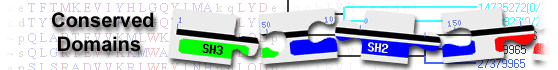
RICIN domain-containing protein( domain architecture ID 12006040)
RICIN domain-containing protein may have carbohydrate-binding function; similar to Mycobacterium tuberculosis protein Rv1419
List of domain hits
| Name | Accession | Description | Interval | E-value | |||
| Ricin_B_lectin | pfam00652 | Ricin-type beta-trefoil lectin domain; |
36-153 | 1.72e-17 | |||
Ricin-type beta-trefoil lectin domain; : Pssm-ID: 395527 [Multi-domain] Cd Length: 126 Bit Score: 73.72 E-value: 1.72e-17
|
|||||||
| Name | Accession | Description | Interval | E-value | |||
| Ricin_B_lectin | pfam00652 | Ricin-type beta-trefoil lectin domain; |
36-153 | 1.72e-17 | |||
Ricin-type beta-trefoil lectin domain; Pssm-ID: 395527 [Multi-domain] Cd Length: 126 Bit Score: 73.72 E-value: 1.72e-17
|
|||||||
| RICIN | smart00458 | Ricin-type beta-trefoil; Carbohydrate-binding domain formed from presumed gene triplication. |
41-156 | 1.88e-15 | |||
Ricin-type beta-trefoil; Carbohydrate-binding domain formed from presumed gene triplication. Pssm-ID: 214672 [Multi-domain] Cd Length: 118 Bit Score: 67.92 E-value: 1.88e-15
|
|||||||
| beta-trefoil_Ricin-like | cd00161 | ricin B-type lectin domain, beta-trefoil fold; The ricin B-type lectin domain is a ... |
35-153 | 8.76e-12 | |||
ricin B-type lectin domain, beta-trefoil fold; The ricin B-type lectin domain is a carbohydrate-binding domain formed from presumed gene triplication. It shows a beta-trefoil fold characterized by 12 beta strands folded into three similar trefoil subdomains (alpha, beta, and gamma) associated to give an overall structure with pseudo-3-fold symmetry. The ricin B-type lectin domain was originally found in Ricin, which is a legume lectin from the seeds of the castor bean plant, Ricinus communis. It is also found in many carbohydrate-recognition proteins like plant and bacterial AB-toxins, glycosidases, or proteases, which serve diverse functions such as inhibitory toxicity, enzymatic activity, and signal transduction. The ricin B-type lectin domain can be present in one or more copies and has been shown in some instances to bind simple sugars, such as galactose or lactose. The most characteristic, though not completely conserved, sequence feature is the presence of a Q-W pattern. Consequently, the ricin B-type lectin domain has also been referred as the (QxW)3 domain and the three homologous regions as the QxW repeats. A disulfide bond is also conserved in some QxW repeats. Pssm-ID: 467293 [Multi-domain] Cd Length: 134 Bit Score: 58.92 E-value: 8.76e-12
|
|||||||
| beta-trefoil_Ricin_XLN-like | cd23418 | ricin B-type lectin domain, beta-trefoil fold, found in Streptomyces olivaceoviridis endo-1, ... |
38-153 | 5.93e-07 | |||
ricin B-type lectin domain, beta-trefoil fold, found in Streptomyces olivaceoviridis endo-1,4-beta-xylanase and similar proteins; The family includes Streptomyces olivaceoviridis endo-1,4-beta-xylanase (XLN, EC 3.2.1.8), Streptomyces avermitilis beta-L-arabinopyranosidase (EC 3.2.1.185), and Streptomyces coelicolor extracellular exo-alpha-L-arabinofuranosidase (ABF, EC 3.2.1.55). XLN, also called xylanase, or 1,4-beta-D-xylan xylanohydrolase, belongs to the glycosyl hydrolase 10 (cellulase F) family. It contributes to hydrolyze hemicellulose, the major component of plant cell walls. XLN contains a (beta/alpha)8-barrel as a catalytic domain, a family 13 carbohydrate binding module (CBM13) as a xylan binding domain (XBD) and a Gly/Pro-rich linker between them. Beta-L-arabinopyranosidase belongs to the glycosyl hydrolase 27 (GH27) family. It has a GH27 catalytic domain, an antiparallel beta-domain containing Greek key motifs, another antiparallel beta-domain forming a jellyroll structure, and a CBM13 module. ScAraf62A, also called ABF, or arabinosidase, or arabinoxylan arabinofuranohydrolase, belongs to the glycosyl hydrolase 62 (GH62) family. It is involved in the degradation of xylan and is a key enzyme in the complete degradation of the plant cell wall. It has a specific arabinofuranose-debranching activity on xylan from gramineae. It acts synergistically with xylanases and binds specifically to xylan. ScAraf62A comprises a CBM13 module at its N-terminus and a catalytic domain at its C-terminus. This model corresponds to the CBM13 module, which is a ricin B-type lectin domain with a beta-trefoil fold, characterized by 12 beta strands folded into three similar trefoil subdomains (alpha, beta, and gamma) associated to give an overall structure with pseudo-3-fold symmetry. Each subdomain may contain a potential sugar-binding pocket. Pssm-ID: 467297 [Multi-domain] Cd Length: 130 Bit Score: 45.80 E-value: 5.93e-07
|
|||||||
| beta-trefoil_Ricin_SCDase | cd23456 | ricin B-type lectin domain, beta-trefoil fold, found in Shewanella algae sphingolipid ceramide ... |
37-112 | 3.85e-06 | |||
ricin B-type lectin domain, beta-trefoil fold, found in Shewanella algae sphingolipid ceramide N-deacylase (SCDase) and similar proteins; SCDase (EC 3.5.1.69) is an enzyme which hydrolyzes the N-acyl linkage between fatty acid and sphingosine in ceramide of various glycosphingolipids and sphingomyelin. Shewanella algae SCDase contains two ricin B-type lectin domains at its C-terminus. The ricin B-type lectin domain shows a beta-trefoil fold, which is characterized by 12 beta strands folded into three similar trefoil subdomains (alpha, beta, and gamma) associated to give an overall structure with pseudo-3-fold symmetry. Each subdomain may harbor a sugar-binding pocket. Pssm-ID: 467334 [Multi-domain] Cd Length: 122 Bit Score: 43.50 E-value: 3.85e-06
|
|||||||
| beta-trefoil_Ricin_GALNT14-like | cd23441 | ricin B-type lectin domain, beta-trefoil fold, found in the polypeptide ... |
38-154 | 4.63e-06 | |||
ricin B-type lectin domain, beta-trefoil fold, found in the polypeptide N-acetylgalactosaminyltransferase 14 (GALNT14)-like subfamily; The GALNT14-like subfamily includes GALNT14 and GALNT16. They act as GalNAc transferases (EC 2.4.1.41) that catalyze the initial reaction in O-linked oligosaccharide biosynthesis, the transfer of an N-acetyl-D-galactosamine residue to a serine or threonine residue on the protein receptor. GALNT14 displays activity toward mucin-derived peptide substrates such as Muc2, Muc5AC, Muc7, and Muc13 (-58). It may be involved in O-glycosylation in the kidney. Members of this subfamily comprise a glycosyltransferase region and a ricin B-type lectin domain. The glycosyltransferase region contains two conserved domains, domain A (also called GT1 motif) and domain B (also called Gal/GalNAc-T motif). This model corresponds to the ricin B-type lectin domain with a beta-trefoil fold, which is characterized by 12 beta strands folded into three similar trefoil subdomains (alpha, beta, and gamma) associated to give an overall structure with pseudo-3-fold symmetry. The ricin B-type lectin domain binds to GalNAc and contributes to the glycopeptide specificity. Pssm-ID: 467319 [Multi-domain] Cd Length: 122 Bit Score: 43.54 E-value: 4.63e-06
|
|||||||
| beta-trefoil_Ricin_AglA | cd23425 | ricin B-type lectin domain, beta-trefoil fold, found in alpha-galactosidase A (AglA) and ... |
63-153 | 5.35e-06 | |||
ricin B-type lectin domain, beta-trefoil fold, found in alpha-galactosidase A (AglA) and similar proteins; AglA (EC 3.2.1.22), also called melibiase A, hydrolyzes a variety of simple alpha-D-galactosides as well as more complex molecules such as oligosaccharides and polysaccharides. It belongs to the glycosyl hydrolase 27 (GH27) family. AglA contains an N-terminal GH27 catalytic domain, an alpha galactosidase C-terminal beta sandwich domain in the middle region, and a carbohydrate-binding domain at the C-terminus. The carbohydrate-binding domain is also known as a ricin B-type lectin domain with a beta-trefoil fold, which is characterized by 12 beta strands folded into three similar trefoil subdomains (alpha, beta, and gamma) associated to give an overall structure with pseudo-3-fold symmetry. Each subdomain may contain a potential sugar-binding pocket. Pssm-ID: 467303 [Multi-domain] Cd Length: 116 Bit Score: 43.20 E-value: 5.35e-06
|
|||||||
| beta-trefoil_Ricin_laminarinase | cd23451 | ricin B-type lectin domain, beta-trefoil fold, found in glucan endo-1,3-beta-glucosidase and ... |
47-153 | 1.26e-05 | |||
ricin B-type lectin domain, beta-trefoil fold, found in glucan endo-1,3-beta-glucosidase and similar proteins; Glucan endo-1,3-beta-glucosidase (EC 3.2.1.39), also called (1->3)-beta-glucan endohydrolase, or (1->3)-beta-glucanase, or laminarinase, belongs to the glycosyl hydrolase 64 (GH64) family. It catalyzes hydrolysis of (1->3)-beta-D-glucosidic linkages in (1->3)-beta-D-glucans. It has been implicated in the defense against fungal pathogens. Glucan endo-1,3-beta-glucosidase contains a C-terminal ricin B-type lectin domain with a beta-trefoil fold, characterized by 12 beta strands folded into three similar trefoil subdomains (alpha, beta, and gamma) associated to give an overall structure with pseudo-3-fold symmetry. Each subdomain bears a potential sugar binding site. Pssm-ID: 467329 [Multi-domain] Cd Length: 125 Bit Score: 42.32 E-value: 1.26e-05
|
|||||||
| beta-trefoil_Ricin_GALNT2 | cd23434 | ricin B-type lectin domain, beta-trefoil fold, found in polypeptide ... |
44-154 | 5.41e-05 | |||
ricin B-type lectin domain, beta-trefoil fold, found in polypeptide N-acetylgalactosaminyltransferase 2 (GALNT2) and similar proteins; GALNT2 (EC 2.4.1.41), also called polypeptide GalNAc transferase 2, GalNAc-T2, pp-GaNTase 2, protein-UDP acetylgalactosaminyltransferase 2, or UDP-GalNAc:polypeptide N-acetylgalactosaminyltransferase 2, catalyzes the initial reaction in O-linked oligosaccharide biosynthesis, the transfer of an N-acetyl-D-galactosamine residue to a serine or threonine residue on the protein receptor. GALNT2 has a broad spectrum of substrates for peptides such as EA2, Muc5AC, Muc1a, Muc1b. It is probably involved in O-linked glycosylation of the immunoglobulin A1 (IgA1) hinge region. It is also involved in O-linked glycosylation of APOC-III, ANGPTL3 and PLTP. It participates in the regulation of HDL-C metabolism. GALNT2 comprises a glycosyltransferase region and a ricin B-type lectin domain. The glycosyltransferase region contains two conserved domains, domain A (also called GT1 motif) and domain B (also called Gal/GalNAc-T motif). This model corresponds to the ricin B-type lectin domain with a beta-trefoil fold, which is characterized by 12 beta strands folded into three similar trefoil subdomains (alpha, beta, and gamma) associated to give an overall structure with pseudo-3-fold symmetry. The ricin B-type lectin domain binds to GalNAc and contributes to the glycopeptide specificity. Pssm-ID: 467312 [Multi-domain] Cd Length: 121 Bit Score: 40.38 E-value: 5.41e-05
|
|||||||
| beta-trefoil_Ricin_XLN-like | cd23418 | ricin B-type lectin domain, beta-trefoil fold, found in Streptomyces olivaceoviridis endo-1, ... |
44-113 | 7.73e-05 | |||
ricin B-type lectin domain, beta-trefoil fold, found in Streptomyces olivaceoviridis endo-1,4-beta-xylanase and similar proteins; The family includes Streptomyces olivaceoviridis endo-1,4-beta-xylanase (XLN, EC 3.2.1.8), Streptomyces avermitilis beta-L-arabinopyranosidase (EC 3.2.1.185), and Streptomyces coelicolor extracellular exo-alpha-L-arabinofuranosidase (ABF, EC 3.2.1.55). XLN, also called xylanase, or 1,4-beta-D-xylan xylanohydrolase, belongs to the glycosyl hydrolase 10 (cellulase F) family. It contributes to hydrolyze hemicellulose, the major component of plant cell walls. XLN contains a (beta/alpha)8-barrel as a catalytic domain, a family 13 carbohydrate binding module (CBM13) as a xylan binding domain (XBD) and a Gly/Pro-rich linker between them. Beta-L-arabinopyranosidase belongs to the glycosyl hydrolase 27 (GH27) family. It has a GH27 catalytic domain, an antiparallel beta-domain containing Greek key motifs, another antiparallel beta-domain forming a jellyroll structure, and a CBM13 module. ScAraf62A, also called ABF, or arabinosidase, or arabinoxylan arabinofuranohydrolase, belongs to the glycosyl hydrolase 62 (GH62) family. It is involved in the degradation of xylan and is a key enzyme in the complete degradation of the plant cell wall. It has a specific arabinofuranose-debranching activity on xylan from gramineae. It acts synergistically with xylanases and binds specifically to xylan. ScAraf62A comprises a CBM13 module at its N-terminus and a catalytic domain at its C-terminus. This model corresponds to the CBM13 module, which is a ricin B-type lectin domain with a beta-trefoil fold, characterized by 12 beta strands folded into three similar trefoil subdomains (alpha, beta, and gamma) associated to give an overall structure with pseudo-3-fold symmetry. Each subdomain may contain a potential sugar-binding pocket. Pssm-ID: 467297 [Multi-domain] Cd Length: 130 Bit Score: 40.03 E-value: 7.73e-05
|
|||||||
| beta-trefoil_Ricin-like | cd00161 | ricin B-type lectin domain, beta-trefoil fold; The ricin B-type lectin domain is a ... |
33-111 | 2.12e-04 | |||
ricin B-type lectin domain, beta-trefoil fold; The ricin B-type lectin domain is a carbohydrate-binding domain formed from presumed gene triplication. It shows a beta-trefoil fold characterized by 12 beta strands folded into three similar trefoil subdomains (alpha, beta, and gamma) associated to give an overall structure with pseudo-3-fold symmetry. The ricin B-type lectin domain was originally found in Ricin, which is a legume lectin from the seeds of the castor bean plant, Ricinus communis. It is also found in many carbohydrate-recognition proteins like plant and bacterial AB-toxins, glycosidases, or proteases, which serve diverse functions such as inhibitory toxicity, enzymatic activity, and signal transduction. The ricin B-type lectin domain can be present in one or more copies and has been shown in some instances to bind simple sugars, such as galactose or lactose. The most characteristic, though not completely conserved, sequence feature is the presence of a Q-W pattern. Consequently, the ricin B-type lectin domain has also been referred as the (QxW)3 domain and the three homologous regions as the QxW repeats. A disulfide bond is also conserved in some QxW repeats. Pssm-ID: 467293 [Multi-domain] Cd Length: 134 Bit Score: 38.89 E-value: 2.12e-04
|
|||||||
| beta-trefoil_Ricin_AH | cd23415 | ricin B-type lectin domain, beta-trefoil fold, found in Actinomycete actinohivin and similar ... |
35-153 | 1.31e-03 | |||
ricin B-type lectin domain, beta-trefoil fold, found in Actinomycete actinohivin and similar proteins; Actinohivin is an actinomycete lectin with a potent specific anti-human immunodeficiency virus (anti-HIV) activity. It inhibits viral entry to cells by binding the high-mannose type sugar chains of gp120. Actinohivin contains a ricin B-type lectin domain with a beta-trefoil fold, which is characterized by 12 beta strands folded into three similar trefoil subdomains (alpha, beta, and gamma) associated to give an overall structure with pseudo-3-fold symmetry. Each subdomain contains a sugar-binding pocket. Pssm-ID: 467294 [Multi-domain] Cd Length: 120 Bit Score: 36.64 E-value: 1.31e-03
|
|||||||
| beta-trefoil_Ricin_CRYBG | cd23430 | ricin B-type lectin domain, beta-trefoil fold, found in the beta/gamma crystallin ... |
108-153 | 1.53e-03 | |||
ricin B-type lectin domain, beta-trefoil fold, found in the beta/gamma crystallin domain-containing protein (CRYBG) family; The CRYBG family includes three members: CRYBG1, CRYBG2, and CRYBG3/vlAKAP. CRYBG1, also called absent in melanoma 1 protein (AIM1), may function as a suppressor of malignant melanoma. It may exert its effects through interactions with the cytoskeleton. CRYBG2 is also called absent in melanoma 1-like protein (AIM1L). CRYBG3/vlAKAP, also called very large A-kinase anchor protein, is an anchoring protein that mediates the subcellular compartmentation of protein kinase A (PKA). It binds to the dimeric RII-alpha regulatory subunit of PKA (PRKAR2A/PRKAR2B). CRYBG proteins belong to the beta/gamma-crystallin family. They all contain a ricin B-type lectin domain with a beta-trefoil fold at the C-terminus. The beta-trefoil fold is characterized by 12 beta strands folded into three similar trefoil subdomains (alpha, beta, and gamma) associated to give an overall structure with pseudo-3-fold symmetry. Each subdomain may harbor a sugar-binding pocket. Pssm-ID: 467308 [Multi-domain] Cd Length: 133 Bit Score: 36.80 E-value: 1.53e-03
|
|||||||
| beta-trefoil_Ricin-like | cd00161 | ricin B-type lectin domain, beta-trefoil fold; The ricin B-type lectin domain is a ... |
31-72 | 1.92e-03 | |||
ricin B-type lectin domain, beta-trefoil fold; The ricin B-type lectin domain is a carbohydrate-binding domain formed from presumed gene triplication. It shows a beta-trefoil fold characterized by 12 beta strands folded into three similar trefoil subdomains (alpha, beta, and gamma) associated to give an overall structure with pseudo-3-fold symmetry. The ricin B-type lectin domain was originally found in Ricin, which is a legume lectin from the seeds of the castor bean plant, Ricinus communis. It is also found in many carbohydrate-recognition proteins like plant and bacterial AB-toxins, glycosidases, or proteases, which serve diverse functions such as inhibitory toxicity, enzymatic activity, and signal transduction. The ricin B-type lectin domain can be present in one or more copies and has been shown in some instances to bind simple sugars, such as galactose or lactose. The most characteristic, though not completely conserved, sequence feature is the presence of a Q-W pattern. Consequently, the ricin B-type lectin domain has also been referred as the (QxW)3 domain and the three homologous regions as the QxW repeats. A disulfide bond is also conserved in some QxW repeats. Pssm-ID: 467293 [Multi-domain] Cd Length: 134 Bit Score: 36.58 E-value: 1.92e-03
|
|||||||
| beta-trefoil_Ricin_Pgant3-like | cd23460 | ricin B-type lectin domain, beta-trefoil fold, found in Drosophila melanogaster polypeptide ... |
37-153 | 6.90e-03 | |||
ricin B-type lectin domain, beta-trefoil fold, found in Drosophila melanogaster polypeptide N-acetylgalactosaminyltransferase 3 (Pgant3) and similar proteins; Pgant3, also called pp-GaNTase 3, protein-UDP acetylgalactosaminyltransferase 3, or UDP-GalNAc:polypeptide N-acetylgalactosaminyltransferase 3, functions as a GalNAc transferase (EC 2.4.1.41) that catalyzes the initial reaction in O-linked oligosaccharide biosynthesis, the transfer of an N-acetyl-D-galactosamine residue to a serine or threonine residue on the protein receptor. Pgant3 can both act as a peptide transferase that transfers GalNAc onto unmodified peptide substrates, and as a glycopeptide transferase that requires the prior addition of a GalNAc on a peptide before adding additional GalNAc moieties. It prefers EA2 as a substrate and has weak activity toward Muc5AC-3, -13 and -3/13 substrates. Pgant3 plays a critical role in the regulation of integrin-mediated cell adhesion during wing development by influencing, via glycosylation, the secretion and localization of the integrin ligand Tig to the basal cell layer interface. It may have a role in protein O-glycosylation in the Golgi and thereby in establishing and/or maintaining a proper secretory apparatus structure. Together with Pgant35A, Pgant3 regulates integrin levels and activity-dependent integrin signaling at the synapse in neurons and muscles. Members of this subfamily contain a ricin B-type lectin domain at the C-terminus. The ricin B-type lectin domain shows a beta-trefoil fold, which is characterized by 12 beta strands folded into three similar trefoil subdomains (alpha, beta, and gamma) associated to give an overall structure with pseudo-3-fold symmetry. Each subdomain bears a potential sugar binding site. Pssm-ID: 467338 [Multi-domain] Cd Length: 121 Bit Score: 34.73 E-value: 6.90e-03
|
|||||||
| Blast search parameters | ||||
|
