

fermitin family homolog 1 isoform X2 [Gallus gallus]
Protein Classification

FRMD7 family protein; ezrin/radixin/moesin family protein( domain architecture ID 13019957)
FRMD7 family protein similar to Homo sapiens FERM domain-containing protein 7 (FRMD7) that plays a role in neurite development| ezrin/radixin/moesin (ERM) family protein links the actin cytoskeleton and the plasma membrane to govern membrane structure and organization
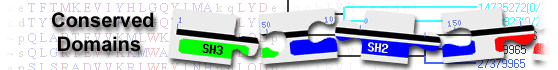
List of domain hits
| Name | Accession | Description | Interval | E-value | ||||
| PH_fermitin | cd01237 | Fermitin family pleckstrin homology (PH) domain; Fermitin functions as a mediator of integrin ... |
370-494 | 3.16e-70 | ||||
Fermitin family pleckstrin homology (PH) domain; Fermitin functions as a mediator of integrin inside-out signalling. The recruitment of Fermitin proteins and Talin to the membrane mediates the terminal event of integrin signalling, via interaction with integrin beta subunits. Fermatin has FERM domain interrupted with a pleckstrin homology (PH) domain. Fermitin family homologs (Fermt1, 2, and 3, also known as Kindlins) are each encoded by a different gene. In mammalian studies, Fermt1 is generally expressed in epithelial cells, Fermt2 is expressed inmuscle tissues, and Fermt3 is expressed in hematopoietic lineages. Specifically Fermt2 is expressed in smooth and striated muscle tissues in mice and in the somites (a trunk muscle precursor) and neural crest in Xenopus embryos. As such it has been proposed that Fermt2 plays a role in cardiomyocyte and neural crest differentiation. Expression of mammalian Fermt3 is associated with hematopoietic lineages: the anterior ventral blood islands, vitelline veins, and early myeloid cells. In Xenopus embryos this expression, also include the notochord and cement gland. PH domains have diverse functions, but in general are involved in targeting proteins to the appropriate cellular location or in the interaction with a binding partner. They share little sequence conservation, but all have a common fold, which is electrostatically polarized. Less than 10% of PH domains bind phosphoinositide phosphates (PIPs) with high affinity and specificity. PH domains are distinguished from other PIP-binding domains by their specific high-affinity binding to PIPs with two vicinal phosphate groups: PtdIns(3,4)P2, PtdIns(4,5)P2 or PtdIns(3,4,5)P3 which results in targeting some PH domain proteins to the plasma membrane. A few display strong specificity in lipid binding. Any specificity is usually determined by loop regions or insertions in the N-terminus of the domain, which are not conserved across all PH domains. PH domains are found in cellular signaling proteins such as serine/threonine kinase, tyrosine kinases, regulators of G-proteins, endocytotic GTPases, adaptors, as well as cytoskeletal associated molecules and in lipid associated enzymes. : Pssm-ID: 269943 Cd Length: 125 Bit Score: 224.19 E-value: 3.16e-70
|
||||||||
| FERM_C_fermitin | cd13205 | FERM domain C-lobe of the Fermitin family; Fermitin functions as a mediator of integrin ... |
564-655 | 1.92e-56 | ||||
FERM domain C-lobe of the Fermitin family; Fermitin functions as a mediator of integrin inside-out signalling. The recruitment of Fermitin proteins and Talin to the membrane mediates the terminal event of integrin signalling, via interaction with integrin beta subunits. Fermatin has FERM domain interrupted with a pleckstrin homology (PH) domain. Fermitin family homologs (Fermt1, 2, and 3, also known as Kindlins) are each encoded by a different gene. In mammalian studies, Fermt1 is generally expressed in epithelial cells, Fermt2 is expressed inmuscle tissues, and Fermt3 is expressed in hematopoietic lineages. Specifically Fermt2 is expressed in smooth and striated muscle tissues in mice and in the somites (a trunk muscle precursor) and neural crest in Xenopus embryos. As such it has been proposed that Fermt2 plays a role in cardiomyocyte and neural crest differentiation. Expression of mammalian Fermt3 is associated with hematopoietic lineages: the anterior ventral blood islands, vitelline veins, and early myeloid cells. In Xenopus embryos this expression, also include the notochord and cement gland. The FERM domain has a cloverleaf tripart structure composed of: (1) FERM_N (A-lobe or F1); (2) FERM_M (B-lobe, or F2); and (3) FERM_C (C-lobe or F3). This cd is not included in the C-lobe hierarchy based on its position in the tree. One thing to note is that unlike the other members of the C-lobe hierarchy it contains 2 FERM M domains which might also reflect a difference in its evolutionary history. The C-lobe/F3 within the FERM domain is part of the PH domain family. The FERM domain is found in the cytoskeletal-associated proteins such as ezrin, moesin, radixin, 4.1R, and merlin. These proteins provide a link between the membrane and cytoskeleton and are involved in signal transduction pathways. The FERM domain is also found in protein tyrosine phosphatases (PTPs), the tyrosine kinases FAK and JAK, in addition to other proteins involved in signaling. This domain is structurally similar to the PH and PTB domains and consequently is capable of binding to both peptides and phospholipids at different sites. : Pssm-ID: 270026 Cd Length: 91 Bit Score: 186.39 E-value: 1.92e-56
|
||||||||
| FERM_F0_KIND1 | cd17180 | FERM (Four.1 protein, Ezrin, Radixin, Moesin) domain, F0 sub-domain, found in kindlin-1 (KIND1) ... |
11-94 | 3.59e-55 | ||||
FERM (Four.1 protein, Ezrin, Radixin, Moesin) domain, F0 sub-domain, found in kindlin-1 (KIND1); KIND1, also termed Kindlerin, or Kindler syndrome protein, or fermitin family homolog 1 (FERMT1), or Unc-112-related protein 1 (URP1), is an integrin-interacting protein that has been implicated in cell adhesion, proliferation, polarity, and motility. It is essential for maintaining the structure of cell-matrix adhesion, such as focal adhesions and podosomes. KIND1 is expressed primarily in epithelial cells. Loss or mutations of KIND1 gene may cause the Kindler syndrome (KS), an autosomal recessive skin disorder with an intriguing progressive phenotype comprising skin blistering, photosensitivity, progressive poikiloderma with extensive skin atrophy, and propensity to skin cancer. KIND1 forms a molecular complex with the key transforming growth factor (TGF)-beta/Smad3 signaling components including type I TGFbeta receptor (TbetaRI), Smad3 and Smad anchor for receptor activation (SARA) to control the activation of TGF-beta/Smad3 signaling pathway. KIND1 consists of an atypical FERM domain that is made up of F1, F2 and F3 domains, as well as an N-terminal region, which precedes the FERM domain and has been referred to as the F0 domain. This family corresponds to the F0 domain. : Pssm-ID: 340700 Cd Length: 84 Bit Score: 182.74 E-value: 3.59e-55
|
||||||||
| FERM_F1_KIND1 | cd17183 | FERM (Four.1 protein, Ezrin, Radixin, Moesin) domain, F1 sub-domain, found in kindlin-1 (KIND1) ... |
95-275 | 1.62e-43 | ||||
FERM (Four.1 protein, Ezrin, Radixin, Moesin) domain, F1 sub-domain, found in kindlin-1 (KIND1); KIND1, also termed Kindlerin, or Kindler syndrome protein, or fermitin family homolog 1 (FERMT1), or Unc-112-related protein 1 (URP1), is an integrin-interacting protein that has been implicated in cell adhesion, proliferation, polarity, and motility. It is essential for maintaining the structure of cell-matrix adhesion, such as focal adhesions and podosomes. KIND1 is expressed primarily in epithelial cells. Loss or mutations of KIND1 gene may cause the Kindler syndrome (KS), an autosomal recessive skin disorder with an intriguing progressive phenotype comprising skin blistering, photosensitivity, progressive poikiloderma with extensive skin atrophy, and propensity to skin cancer. KIND1 forms a molecular complex with the key transforming growth factor (TGF)-beta/Smad3 signaling components including type I TGFbeta receptor (TbetaRI), Smad3 and Smad anchor for receptor activation (SARA) to control the activation of TGF-beta/Smad3 signaling pathway. KIND1 consists of an atypical FERM domain that is made up of F1, F2 and F3 domains, as well as an N-terminal region, which precedes the FERM domain and has been referred to as the F0 domain. This family corresponds to the F1 domain. : Pssm-ID: 340703 Cd Length: 93 Bit Score: 151.14 E-value: 1.62e-43
|
||||||||
| B41 | smart00295 | Band 4.1 homologues; Also known as ezrin/radixin/moesin (ERM) protein domains. Present in ... |
250-337 | 1.49e-18 | ||||
Band 4.1 homologues; Also known as ezrin/radixin/moesin (ERM) protein domains. Present in myosins, ezrin, radixin, moesin, protein tyrosine phosphatases. Plasma membrane-binding domain. These proteins play structural and regulatory roles in the assembly and stabilization of specialized plasmamembrane domains. Some PDZ domain containing proteins bind one or more of this family. Now includes JAKs. : Pssm-ID: 214604 [Multi-domain] Cd Length: 201 Bit Score: 84.27 E-value: 1.49e-18
|
||||||||
| FERM_M | pfam00373 | FERM central domain; This domain is the central structural domain of the FERM domain. |
453-570 | 3.37e-14 | ||||
FERM central domain; This domain is the central structural domain of the FERM domain. : Pssm-ID: 459788 [Multi-domain] Cd Length: 117 Bit Score: 69.22 E-value: 3.37e-14
|
||||||||
| Name | Accession | Description | Interval | E-value | ||||
| PH_fermitin | cd01237 | Fermitin family pleckstrin homology (PH) domain; Fermitin functions as a mediator of integrin ... |
370-494 | 3.16e-70 | ||||
Fermitin family pleckstrin homology (PH) domain; Fermitin functions as a mediator of integrin inside-out signalling. The recruitment of Fermitin proteins and Talin to the membrane mediates the terminal event of integrin signalling, via interaction with integrin beta subunits. Fermatin has FERM domain interrupted with a pleckstrin homology (PH) domain. Fermitin family homologs (Fermt1, 2, and 3, also known as Kindlins) are each encoded by a different gene. In mammalian studies, Fermt1 is generally expressed in epithelial cells, Fermt2 is expressed inmuscle tissues, and Fermt3 is expressed in hematopoietic lineages. Specifically Fermt2 is expressed in smooth and striated muscle tissues in mice and in the somites (a trunk muscle precursor) and neural crest in Xenopus embryos. As such it has been proposed that Fermt2 plays a role in cardiomyocyte and neural crest differentiation. Expression of mammalian Fermt3 is associated with hematopoietic lineages: the anterior ventral blood islands, vitelline veins, and early myeloid cells. In Xenopus embryos this expression, also include the notochord and cement gland. PH domains have diverse functions, but in general are involved in targeting proteins to the appropriate cellular location or in the interaction with a binding partner. They share little sequence conservation, but all have a common fold, which is electrostatically polarized. Less than 10% of PH domains bind phosphoinositide phosphates (PIPs) with high affinity and specificity. PH domains are distinguished from other PIP-binding domains by their specific high-affinity binding to PIPs with two vicinal phosphate groups: PtdIns(3,4)P2, PtdIns(4,5)P2 or PtdIns(3,4,5)P3 which results in targeting some PH domain proteins to the plasma membrane. A few display strong specificity in lipid binding. Any specificity is usually determined by loop regions or insertions in the N-terminus of the domain, which are not conserved across all PH domains. PH domains are found in cellular signaling proteins such as serine/threonine kinase, tyrosine kinases, regulators of G-proteins, endocytotic GTPases, adaptors, as well as cytoskeletal associated molecules and in lipid associated enzymes. Pssm-ID: 269943 Cd Length: 125 Bit Score: 224.19 E-value: 3.16e-70
|
||||||||
| FERM_C_fermitin | cd13205 | FERM domain C-lobe of the Fermitin family; Fermitin functions as a mediator of integrin ... |
564-655 | 1.92e-56 | ||||
FERM domain C-lobe of the Fermitin family; Fermitin functions as a mediator of integrin inside-out signalling. The recruitment of Fermitin proteins and Talin to the membrane mediates the terminal event of integrin signalling, via interaction with integrin beta subunits. Fermatin has FERM domain interrupted with a pleckstrin homology (PH) domain. Fermitin family homologs (Fermt1, 2, and 3, also known as Kindlins) are each encoded by a different gene. In mammalian studies, Fermt1 is generally expressed in epithelial cells, Fermt2 is expressed inmuscle tissues, and Fermt3 is expressed in hematopoietic lineages. Specifically Fermt2 is expressed in smooth and striated muscle tissues in mice and in the somites (a trunk muscle precursor) and neural crest in Xenopus embryos. As such it has been proposed that Fermt2 plays a role in cardiomyocyte and neural crest differentiation. Expression of mammalian Fermt3 is associated with hematopoietic lineages: the anterior ventral blood islands, vitelline veins, and early myeloid cells. In Xenopus embryos this expression, also include the notochord and cement gland. The FERM domain has a cloverleaf tripart structure composed of: (1) FERM_N (A-lobe or F1); (2) FERM_M (B-lobe, or F2); and (3) FERM_C (C-lobe or F3). This cd is not included in the C-lobe hierarchy based on its position in the tree. One thing to note is that unlike the other members of the C-lobe hierarchy it contains 2 FERM M domains which might also reflect a difference in its evolutionary history. The C-lobe/F3 within the FERM domain is part of the PH domain family. The FERM domain is found in the cytoskeletal-associated proteins such as ezrin, moesin, radixin, 4.1R, and merlin. These proteins provide a link between the membrane and cytoskeleton and are involved in signal transduction pathways. The FERM domain is also found in protein tyrosine phosphatases (PTPs), the tyrosine kinases FAK and JAK, in addition to other proteins involved in signaling. This domain is structurally similar to the PH and PTB domains and consequently is capable of binding to both peptides and phospholipids at different sites. Pssm-ID: 270026 Cd Length: 91 Bit Score: 186.39 E-value: 1.92e-56
|
||||||||
| FERM_F0_KIND1 | cd17180 | FERM (Four.1 protein, Ezrin, Radixin, Moesin) domain, F0 sub-domain, found in kindlin-1 (KIND1) ... |
11-94 | 3.59e-55 | ||||
FERM (Four.1 protein, Ezrin, Radixin, Moesin) domain, F0 sub-domain, found in kindlin-1 (KIND1); KIND1, also termed Kindlerin, or Kindler syndrome protein, or fermitin family homolog 1 (FERMT1), or Unc-112-related protein 1 (URP1), is an integrin-interacting protein that has been implicated in cell adhesion, proliferation, polarity, and motility. It is essential for maintaining the structure of cell-matrix adhesion, such as focal adhesions and podosomes. KIND1 is expressed primarily in epithelial cells. Loss or mutations of KIND1 gene may cause the Kindler syndrome (KS), an autosomal recessive skin disorder with an intriguing progressive phenotype comprising skin blistering, photosensitivity, progressive poikiloderma with extensive skin atrophy, and propensity to skin cancer. KIND1 forms a molecular complex with the key transforming growth factor (TGF)-beta/Smad3 signaling components including type I TGFbeta receptor (TbetaRI), Smad3 and Smad anchor for receptor activation (SARA) to control the activation of TGF-beta/Smad3 signaling pathway. KIND1 consists of an atypical FERM domain that is made up of F1, F2 and F3 domains, as well as an N-terminal region, which precedes the FERM domain and has been referred to as the F0 domain. This family corresponds to the F0 domain. Pssm-ID: 340700 Cd Length: 84 Bit Score: 182.74 E-value: 3.59e-55
|
||||||||
| FERM_F1_KIND1 | cd17183 | FERM (Four.1 protein, Ezrin, Radixin, Moesin) domain, F1 sub-domain, found in kindlin-1 (KIND1) ... |
95-275 | 1.62e-43 | ||||
FERM (Four.1 protein, Ezrin, Radixin, Moesin) domain, F1 sub-domain, found in kindlin-1 (KIND1); KIND1, also termed Kindlerin, or Kindler syndrome protein, or fermitin family homolog 1 (FERMT1), or Unc-112-related protein 1 (URP1), is an integrin-interacting protein that has been implicated in cell adhesion, proliferation, polarity, and motility. It is essential for maintaining the structure of cell-matrix adhesion, such as focal adhesions and podosomes. KIND1 is expressed primarily in epithelial cells. Loss or mutations of KIND1 gene may cause the Kindler syndrome (KS), an autosomal recessive skin disorder with an intriguing progressive phenotype comprising skin blistering, photosensitivity, progressive poikiloderma with extensive skin atrophy, and propensity to skin cancer. KIND1 forms a molecular complex with the key transforming growth factor (TGF)-beta/Smad3 signaling components including type I TGFbeta receptor (TbetaRI), Smad3 and Smad anchor for receptor activation (SARA) to control the activation of TGF-beta/Smad3 signaling pathway. KIND1 consists of an atypical FERM domain that is made up of F1, F2 and F3 domains, as well as an N-terminal region, which precedes the FERM domain and has been referred to as the F0 domain. This family corresponds to the F1 domain. Pssm-ID: 340703 Cd Length: 93 Bit Score: 151.14 E-value: 1.62e-43
|
||||||||
| Kindlin_2_N | pfam18124 | Kindlin-2 N-terminal domain; This is the N-terminal domain (K2-N) of Kindlin-2 protein present ... |
7-95 | 8.37e-40 | ||||
Kindlin-2 N-terminal domain; This is the N-terminal domain (K2-N) of Kindlin-2 protein present in Homo sapiens. Kindlin-2 is a regulator for heterodimeric integrin adhesion receptors promotes integrin activation. Activation depends on binding of the N-terminal domain to the integrin beta cytoplasmic tail (CT), which disrupts the receptors association with alpha-CT and triggers the conformational transitions in the receptor. K2-N contains a conserved positively charged surface that binds to membrane enriched with negatively charged phosphatidylinositol-(4,5)-bisphosphate (PIP2). K2-N is also very similar to the homologous kindlin-1 F0. Pssm-ID: 465660 Cd Length: 89 Bit Score: 140.85 E-value: 8.37e-40
|
||||||||
| B41 | smart00295 | Band 4.1 homologues; Also known as ezrin/radixin/moesin (ERM) protein domains. Present in ... |
250-337 | 1.49e-18 | ||||
Band 4.1 homologues; Also known as ezrin/radixin/moesin (ERM) protein domains. Present in myosins, ezrin, radixin, moesin, protein tyrosine phosphatases. Plasma membrane-binding domain. These proteins play structural and regulatory roles in the assembly and stabilization of specialized plasmamembrane domains. Some PDZ domain containing proteins bind one or more of this family. Now includes JAKs. Pssm-ID: 214604 [Multi-domain] Cd Length: 201 Bit Score: 84.27 E-value: 1.49e-18
|
||||||||
| FERM_M | pfam00373 | FERM central domain; This domain is the central structural domain of the FERM domain. |
453-570 | 3.37e-14 | ||||
FERM central domain; This domain is the central structural domain of the FERM domain. Pssm-ID: 459788 [Multi-domain] Cd Length: 117 Bit Score: 69.22 E-value: 3.37e-14
|
||||||||
| PH | pfam00169 | PH domain; PH stands for pleckstrin homology. |
390-467 | 3.77e-10 | ||||
PH domain; PH stands for pleckstrin homology. Pssm-ID: 459697 [Multi-domain] Cd Length: 105 Bit Score: 57.57 E-value: 3.77e-10
|
||||||||
| PH | smart00233 | Pleckstrin homology domain; Domain commonly found in eukaryotic signalling proteins. The ... |
378-467 | 8.09e-09 | ||||
Pleckstrin homology domain; Domain commonly found in eukaryotic signalling proteins. The domain family possesses multiple functions including the abilities to bind inositol phosphates, and various proteins. PH domains have been found to possess inserted domains (such as in PLC gamma, syntrophins) and to be inserted within other domains. Mutations in Brutons tyrosine kinase (Btk) within its PH domain cause X-linked agammaglobulinaemia (XLA) in patients. Point mutations cluster into the positively charged end of the molecule around the predicted binding site for phosphatidylinositol lipids. Pssm-ID: 214574 [Multi-domain] Cd Length: 102 Bit Score: 53.71 E-value: 8.09e-09
|
||||||||
| Name | Accession | Description | Interval | E-value | ||||
| PH_fermitin | cd01237 | Fermitin family pleckstrin homology (PH) domain; Fermitin functions as a mediator of integrin ... |
370-494 | 3.16e-70 | ||||
Fermitin family pleckstrin homology (PH) domain; Fermitin functions as a mediator of integrin inside-out signalling. The recruitment of Fermitin proteins and Talin to the membrane mediates the terminal event of integrin signalling, via interaction with integrin beta subunits. Fermatin has FERM domain interrupted with a pleckstrin homology (PH) domain. Fermitin family homologs (Fermt1, 2, and 3, also known as Kindlins) are each encoded by a different gene. In mammalian studies, Fermt1 is generally expressed in epithelial cells, Fermt2 is expressed inmuscle tissues, and Fermt3 is expressed in hematopoietic lineages. Specifically Fermt2 is expressed in smooth and striated muscle tissues in mice and in the somites (a trunk muscle precursor) and neural crest in Xenopus embryos. As such it has been proposed that Fermt2 plays a role in cardiomyocyte and neural crest differentiation. Expression of mammalian Fermt3 is associated with hematopoietic lineages: the anterior ventral blood islands, vitelline veins, and early myeloid cells. In Xenopus embryos this expression, also include the notochord and cement gland. PH domains have diverse functions, but in general are involved in targeting proteins to the appropriate cellular location or in the interaction with a binding partner. They share little sequence conservation, but all have a common fold, which is electrostatically polarized. Less than 10% of PH domains bind phosphoinositide phosphates (PIPs) with high affinity and specificity. PH domains are distinguished from other PIP-binding domains by their specific high-affinity binding to PIPs with two vicinal phosphate groups: PtdIns(3,4)P2, PtdIns(4,5)P2 or PtdIns(3,4,5)P3 which results in targeting some PH domain proteins to the plasma membrane. A few display strong specificity in lipid binding. Any specificity is usually determined by loop regions or insertions in the N-terminus of the domain, which are not conserved across all PH domains. PH domains are found in cellular signaling proteins such as serine/threonine kinase, tyrosine kinases, regulators of G-proteins, endocytotic GTPases, adaptors, as well as cytoskeletal associated molecules and in lipid associated enzymes. Pssm-ID: 269943 Cd Length: 125 Bit Score: 224.19 E-value: 3.16e-70
|
||||||||
| FERM_C_fermitin | cd13205 | FERM domain C-lobe of the Fermitin family; Fermitin functions as a mediator of integrin ... |
564-655 | 1.92e-56 | ||||
FERM domain C-lobe of the Fermitin family; Fermitin functions as a mediator of integrin inside-out signalling. The recruitment of Fermitin proteins and Talin to the membrane mediates the terminal event of integrin signalling, via interaction with integrin beta subunits. Fermatin has FERM domain interrupted with a pleckstrin homology (PH) domain. Fermitin family homologs (Fermt1, 2, and 3, also known as Kindlins) are each encoded by a different gene. In mammalian studies, Fermt1 is generally expressed in epithelial cells, Fermt2 is expressed inmuscle tissues, and Fermt3 is expressed in hematopoietic lineages. Specifically Fermt2 is expressed in smooth and striated muscle tissues in mice and in the somites (a trunk muscle precursor) and neural crest in Xenopus embryos. As such it has been proposed that Fermt2 plays a role in cardiomyocyte and neural crest differentiation. Expression of mammalian Fermt3 is associated with hematopoietic lineages: the anterior ventral blood islands, vitelline veins, and early myeloid cells. In Xenopus embryos this expression, also include the notochord and cement gland. The FERM domain has a cloverleaf tripart structure composed of: (1) FERM_N (A-lobe or F1); (2) FERM_M (B-lobe, or F2); and (3) FERM_C (C-lobe or F3). This cd is not included in the C-lobe hierarchy based on its position in the tree. One thing to note is that unlike the other members of the C-lobe hierarchy it contains 2 FERM M domains which might also reflect a difference in its evolutionary history. The C-lobe/F3 within the FERM domain is part of the PH domain family. The FERM domain is found in the cytoskeletal-associated proteins such as ezrin, moesin, radixin, 4.1R, and merlin. These proteins provide a link between the membrane and cytoskeleton and are involved in signal transduction pathways. The FERM domain is also found in protein tyrosine phosphatases (PTPs), the tyrosine kinases FAK and JAK, in addition to other proteins involved in signaling. This domain is structurally similar to the PH and PTB domains and consequently is capable of binding to both peptides and phospholipids at different sites. Pssm-ID: 270026 Cd Length: 91 Bit Score: 186.39 E-value: 1.92e-56
|
||||||||
| FERM_F0_KIND1 | cd17180 | FERM (Four.1 protein, Ezrin, Radixin, Moesin) domain, F0 sub-domain, found in kindlin-1 (KIND1) ... |
11-94 | 3.59e-55 | ||||
FERM (Four.1 protein, Ezrin, Radixin, Moesin) domain, F0 sub-domain, found in kindlin-1 (KIND1); KIND1, also termed Kindlerin, or Kindler syndrome protein, or fermitin family homolog 1 (FERMT1), or Unc-112-related protein 1 (URP1), is an integrin-interacting protein that has been implicated in cell adhesion, proliferation, polarity, and motility. It is essential for maintaining the structure of cell-matrix adhesion, such as focal adhesions and podosomes. KIND1 is expressed primarily in epithelial cells. Loss or mutations of KIND1 gene may cause the Kindler syndrome (KS), an autosomal recessive skin disorder with an intriguing progressive phenotype comprising skin blistering, photosensitivity, progressive poikiloderma with extensive skin atrophy, and propensity to skin cancer. KIND1 forms a molecular complex with the key transforming growth factor (TGF)-beta/Smad3 signaling components including type I TGFbeta receptor (TbetaRI), Smad3 and Smad anchor for receptor activation (SARA) to control the activation of TGF-beta/Smad3 signaling pathway. KIND1 consists of an atypical FERM domain that is made up of F1, F2 and F3 domains, as well as an N-terminal region, which precedes the FERM domain and has been referred to as the F0 domain. This family corresponds to the F0 domain. Pssm-ID: 340700 Cd Length: 84 Bit Score: 182.74 E-value: 3.59e-55
|
||||||||
| FERM_F1_KIND1 | cd17183 | FERM (Four.1 protein, Ezrin, Radixin, Moesin) domain, F1 sub-domain, found in kindlin-1 (KIND1) ... |
95-275 | 1.62e-43 | ||||
FERM (Four.1 protein, Ezrin, Radixin, Moesin) domain, F1 sub-domain, found in kindlin-1 (KIND1); KIND1, also termed Kindlerin, or Kindler syndrome protein, or fermitin family homolog 1 (FERMT1), or Unc-112-related protein 1 (URP1), is an integrin-interacting protein that has been implicated in cell adhesion, proliferation, polarity, and motility. It is essential for maintaining the structure of cell-matrix adhesion, such as focal adhesions and podosomes. KIND1 is expressed primarily in epithelial cells. Loss or mutations of KIND1 gene may cause the Kindler syndrome (KS), an autosomal recessive skin disorder with an intriguing progressive phenotype comprising skin blistering, photosensitivity, progressive poikiloderma with extensive skin atrophy, and propensity to skin cancer. KIND1 forms a molecular complex with the key transforming growth factor (TGF)-beta/Smad3 signaling components including type I TGFbeta receptor (TbetaRI), Smad3 and Smad anchor for receptor activation (SARA) to control the activation of TGF-beta/Smad3 signaling pathway. KIND1 consists of an atypical FERM domain that is made up of F1, F2 and F3 domains, as well as an N-terminal region, which precedes the FERM domain and has been referred to as the F0 domain. This family corresponds to the F1 domain. Pssm-ID: 340703 Cd Length: 93 Bit Score: 151.14 E-value: 1.62e-43
|
||||||||
| FERM_F0_kindlins | cd17095 | FERM (Four.1 protein, Ezrin, Radixin, Moesin) domain, F0 sub-domain, found in the kindlin ... |
11-94 | 1.01e-42 | ||||
FERM (Four.1 protein, Ezrin, Radixin, Moesin) domain, F0 sub-domain, found in the kindlin family; The kindlin family is composed of kindlin-1, 2 and 3, which are FERM domain-containing adaptor molecules that interact with the cytoplasmic component of integrins and regulate cell-matrix connections. Kindlins belong to the 4.1- ezrin-ridixin-moesin (FERM) domain containing protein family. They contain F1, F2 and F3 subdomains that typify FERM family members, and these subdomains are preceded by an N-terminal F0 subdomain. Both F0 and F1 domains have similar ubiquitin-like folds. This family corresponds to the F0 domain. In addition, a distinctive feature of kindlins is the insertion of a pleckstrin homology (PH) subdomain into the F2 subdomain. Pssm-ID: 340615 Cd Length: 80 Bit Score: 148.60 E-value: 1.01e-42
|
||||||||
| FERM_F0_KIND3 | cd17182 | FERM (Four.1 protein, Ezrin, Radixin, Moesin) domain, F0 sub-domain, found in kindlin-3 (KIND3) ... |
11-94 | 1.85e-40 | ||||
FERM (Four.1 protein, Ezrin, Radixin, Moesin) domain, F0 sub-domain, found in kindlin-3 (KIND3); KIND3, also termed fermitin family homolog 3 (FERMT3), or MIG2-like protein, or Unc-112-related protein 2, is an adaptor protein that expressed primarily in hematopoietic cells. It plays a central role in cell adhesion in hematopoietic cells, and also promotes integrin activation, clustering and outside-in signaling. KIND3, together with talin-1, contributes essentially to the activation of beta2-integrins in neutrophils. In addition, KIND3 interacts with the ribosome and regulates c-Myc expression required for proliferation of chronic myeloid leukemia cells. Mutations in the KIND3 gene cause leukocyte adhesion deficiency type III (LAD III), which is characterized by high susceptibility to infections, spontaneous and episodic bleedings, and osteopetrosis. KIND3 consists of an atypical FERM domain that is made up of F1, F2 and F3 domains, as well as an N-terminal region, which precedes the FERM domain and has been referred to as the F0 domain. This family corresponds to the F0 domain. Pssm-ID: 340702 Cd Length: 83 Bit Score: 142.71 E-value: 1.85e-40
|
||||||||
| Kindlin_2_N | pfam18124 | Kindlin-2 N-terminal domain; This is the N-terminal domain (K2-N) of Kindlin-2 protein present ... |
7-95 | 8.37e-40 | ||||
Kindlin-2 N-terminal domain; This is the N-terminal domain (K2-N) of Kindlin-2 protein present in Homo sapiens. Kindlin-2 is a regulator for heterodimeric integrin adhesion receptors promotes integrin activation. Activation depends on binding of the N-terminal domain to the integrin beta cytoplasmic tail (CT), which disrupts the receptors association with alpha-CT and triggers the conformational transitions in the receptor. K2-N contains a conserved positively charged surface that binds to membrane enriched with negatively charged phosphatidylinositol-(4,5)-bisphosphate (PIP2). K2-N is also very similar to the homologous kindlin-1 F0. Pssm-ID: 465660 Cd Length: 89 Bit Score: 140.85 E-value: 8.37e-40
|
||||||||
| FERM_F0_KIND2 | cd17181 | FERM (Four.1 protein, Ezrin, Radixin, Moesin) domain, F0 sub-domain, found in kindlin-2 (KIND2) ... |
11-94 | 4.10e-35 | ||||
FERM (Four.1 protein, Ezrin, Radixin, Moesin) domain, F0 sub-domain, found in kindlin-2 (KIND2); KIND2, also termed fermitin family homolog 2 (FERMT2), or mitogen-inducible gene 2 protein (MIG-2), or Pleckstrin homology (PH) domain-containing family C member 1, is an adaptor protein that is widely distributed and is particularly abundant in adherent cells. It binds to the integrin beta cytoplasmic tail to promote integrin activation. It promotes carcinogenesis through regulation of cell-cell and cell-extracellular matrix adhesion. In additon, KIND2 plays an important role in cardiac development. KIND2 consists of an atypical FERM domain that is made up of F1, F2 and F3 domains, as well as an N-terminal region, which precedes the FERM domain and has been referred to as the F0 domain. This family corresponds to the F0 domain. Pssm-ID: 340701 Cd Length: 80 Bit Score: 127.49 E-value: 4.10e-35
|
||||||||
| FERM_F1_kindlins | cd17096 | FERM (Four.1 protein, Ezrin, Radixin, Moesin) domain, F1 sub-domain, found in the kindlin ... |
95-275 | 3.53e-33 | ||||
FERM (Four.1 protein, Ezrin, Radixin, Moesin) domain, F1 sub-domain, found in the kindlin family; The kindlin family is composed of Kindlin-1, 2 and 3, which are FERM domain-containing adaptor molecules that interact with the cytoplasmic component of integrins and regulate cell-matrix connections. Kindlins belong to the 4.1- ezrin-ridixin-moesin (FERM) domain containing protein family. They contain F1, F2 and F3 subdomains that typify FERM family members, and these subdomains are preceded by an N-terminal F0 subdomain. Both F0 and F1 domains have similar ubiquitin-like folds. This family corresponds to the F1 domain. In addition, a distinctive feature of kindlins is the insertion of a pleckstrin homology (PH) subdomain into the F2 subdomain. Pssm-ID: 340616 Cd Length: 90 Bit Score: 122.39 E-value: 3.53e-33
|
||||||||
| FERM_F1_KIND2 | cd17184 | FERM (Four.1 protein, Ezrin, Radixin, Moesin) domain, F1 sub-domain, found in kindlin-2 (KIND2) ... |
95-275 | 9.92e-31 | ||||
FERM (Four.1 protein, Ezrin, Radixin, Moesin) domain, F1 sub-domain, found in kindlin-2 (KIND2); KIND2, also termed fermitin family homolog 2 (FERMT2), or mitogen-inducible gene 2 protein (MIG-2), or Pleckstrin homology (PH) domain-containing family C member 1, is an adaptor protein that is widely distributed and is particularly abundant in adherent cells. It binds to the integrin beta cytoplasmic tail to promote integrin activation. It promotes carcinogenesis through regulation of cell-cell and cell-extracellular matrix adhesion. KIND2 also plays an important role in cardiac development. KIND2 consists of an atypical FERM domain that is made up of F1, F2 and F3 domains, as well as an N-terminal region, which precedes the FERM domain and has been referred to as the F0 domain. This family corresponds to the F1 domain. Pssm-ID: 340704 Cd Length: 101 Bit Score: 115.89 E-value: 9.92e-31
|
||||||||
| FERM_F1_KIND3 | cd17185 | FERM (Four.1 protein, Ezrin, Radixin, Moesin) domain, F1 sub-domain, found in kindlin-3 (KIND3) ... |
95-275 | 2.40e-21 | ||||
FERM (Four.1 protein, Ezrin, Radixin, Moesin) domain, F1 sub-domain, found in kindlin-3 (KIND3); KIND3, also termed fermitin family homolog 3 (FERMT3), or MIG2-like protein, or Unc-112-related protein 2, is an adaptor protein that expressed primarily in hematopoietic cells. It plays a central role in cell adhesion in hematopoietic cells, and also promotes integrin activation, clustering and outside-in signaling. KIND3, together with talin-1, contributes essentially to the activation of beta2-integrins in neutrophils. In addition, KIND3 interacts with the ribosome and regulates c-Myc expression required for proliferation of chronic myeloid leukemia cells. Mutations in the KIND3 gene cause leukocyte adhesion deficiency type III (LAD III), which is characterized by high susceptibility to infections, spontaneous and episodic bleedings, and osteopetrosis. KIND3 consists of an atypical FERM domain that is made up of F1, F2 and F3 domains, as well as an N-terminal region, which precedes the FERM domain and has been referred to as the F0 domain. This family corresponds to the F1 domain. Pssm-ID: 340705 Cd Length: 91 Bit Score: 88.77 E-value: 2.40e-21
|
||||||||
| B41 | smart00295 | Band 4.1 homologues; Also known as ezrin/radixin/moesin (ERM) protein domains. Present in ... |
250-337 | 1.49e-18 | ||||
Band 4.1 homologues; Also known as ezrin/radixin/moesin (ERM) protein domains. Present in myosins, ezrin, radixin, moesin, protein tyrosine phosphatases. Plasma membrane-binding domain. These proteins play structural and regulatory roles in the assembly and stabilization of specialized plasmamembrane domains. Some PDZ domain containing proteins bind one or more of this family. Now includes JAKs. Pssm-ID: 214604 [Multi-domain] Cd Length: 201 Bit Score: 84.27 E-value: 1.49e-18
|
||||||||
| FERM_M | pfam00373 | FERM central domain; This domain is the central structural domain of the FERM domain. |
453-570 | 3.37e-14 | ||||
FERM central domain; This domain is the central structural domain of the FERM domain. Pssm-ID: 459788 [Multi-domain] Cd Length: 117 Bit Score: 69.22 E-value: 3.37e-14
|
||||||||
| PH | pfam00169 | PH domain; PH stands for pleckstrin homology. |
390-467 | 3.77e-10 | ||||
PH domain; PH stands for pleckstrin homology. Pssm-ID: 459697 [Multi-domain] Cd Length: 105 Bit Score: 57.57 E-value: 3.77e-10
|
||||||||
| FERM_C-lobe | cd00836 | FERM domain C-lobe; The FERM domain has a cloverleaf tripart structure composed of: (1) FERM_N ... |
566-653 | 5.22e-10 | ||||
FERM domain C-lobe; The FERM domain has a cloverleaf tripart structure composed of: (1) FERM_N (A-lobe or F1); (2) FERM_M (B-lobe, or F2); and (3) FERM_C (C-lobe or F3). The C-lobe/F3 within the FERM domain is part of the PH domain family. The FERM domain is found in the cytoskeletal-associated proteins such as ezrin, moesin, radixin, 4.1R, and merlin. These proteins provide a link between the membrane and cytoskeleton and are involved in signal transduction pathways. The FERM domain is also found in protein tyrosine phosphatases (PTPs), the tyrosine kinases FAK and JAK, in addition to other proteins involved in signaling. This domain is structurally similar to the PH and PTB domains and consequently is capable of binding to both peptides and phospholipids at different sites. Pssm-ID: 275389 Cd Length: 93 Bit Score: 56.61 E-value: 5.22e-10
|
||||||||
| PH | smart00233 | Pleckstrin homology domain; Domain commonly found in eukaryotic signalling proteins. The ... |
378-467 | 8.09e-09 | ||||
Pleckstrin homology domain; Domain commonly found in eukaryotic signalling proteins. The domain family possesses multiple functions including the abilities to bind inositol phosphates, and various proteins. PH domains have been found to possess inserted domains (such as in PLC gamma, syntrophins) and to be inserted within other domains. Mutations in Brutons tyrosine kinase (Btk) within its PH domain cause X-linked agammaglobulinaemia (XLA) in patients. Point mutations cluster into the positively charged end of the molecule around the predicted binding site for phosphatidylinositol lipids. Pssm-ID: 214574 [Multi-domain] Cd Length: 102 Bit Score: 53.71 E-value: 8.09e-09
|
||||||||
| PH | cd00821 | Pleckstrin homology (PH) domain; PH domains have diverse functions, but in general are ... |
378-467 | 1.32e-08 | ||||
Pleckstrin homology (PH) domain; PH domains have diverse functions, but in general are involved in targeting proteins to the appropriate cellular location or in the interaction with a binding partner. They share little sequence conservation, but all have a common fold, which is electrostatically polarized. Less than 10% of PH domains bind phosphoinositide phosphates (PIPs) with high affinity and specificity. PH domains are distinguished from other PIP-binding domains by their specific high-affinity binding to PIPs with two vicinal phosphate groups: PtdIns(3,4)P2, PtdIns(4,5)P2 or PtdIns(3,4,5)P3 which results in targeting some PH domain proteins to the plasma membrane. A few display strong specificity in lipid binding. Any specificity is usually determined by loop regions or insertions in the N-terminus of the domain, which are not conserved across all PH domains. PH domains are found in cellular signaling proteins such as serine/threonine kinase, tyrosine kinases, regulators of G-proteins, endocytotic GTPases, adaptors, as well as cytoskeletal associated molecules and in lipid associated enzymes. Pssm-ID: 275388 [Multi-domain] Cd Length: 92 Bit Score: 52.54 E-value: 1.32e-08
|
||||||||
| PH_Boi | cd13316 | Boi family Pleckstrin homology domain; Yeast Boi proteins Boi1 and Boi2 are functionally ... |
374-472 | 4.79e-07 | ||||
Boi family Pleckstrin homology domain; Yeast Boi proteins Boi1 and Boi2 are functionally redundant and important for cell growth with Boi mutants displaying defects in bud formation and in the maintenance of cell polarity.They appear to be linked to Rho-type GTPase, Cdc42 and Rho3. Boi1 and Boi2 display two-hybrid interactions with the GTP-bound ("active") form of Cdc42, while Rho3 can suppress of the lethality caused by deletion of Boi1 and Boi2. These findings suggest that Boi1 and Boi2 are targets of Cdc42 that promote cell growth in a manner that is regulated by Rho3. Boi proteins contain a N-terminal SH3 domain, followed by a SAM (sterile alpha motif) domain, a proline-rich region, which mediates binding to the second SH3 domain of Bem1, and C-terminal PH domain. The PH domain is essential for its function in cell growth and is important for localization to the bud, while the SH3 domain is needed for localization to the neck. PH domains have diverse functions, but in general are involved in targeting proteins to the appropriate cellular location or in the interaction with a binding partner. They share little sequence conservation, but all have a common fold, which is electrostatically polarized. Less than 10% of PH domains bind phosphoinositide phosphates (PIPs) with high affinity and specificity. PH domains are distinguished from other PIP-binding domains by their specific high-affinity binding to PIPs with two vicinal phosphate groups: PtdIns(3,4)P2, PtdIns(4,5)P2 or PtdIns(3,4,5)P3 which results in targeting some PH domain proteins to the plasma membrane. A few display strong specificity in lipid binding. Any specificity is usually determined by loop regions or insertions in the N-terminus of the domain, which are not conserved across all PH domains. PH domains are found in cellular signaling proteins such as serine/threonine kinase, tyrosine kinases, regulators of G-proteins, endocytotic GTPases, adaptors, as well as cytoskeletal associated molecules and in lipid associated enzymes. Pssm-ID: 270126 Cd Length: 97 Bit Score: 48.14 E-value: 4.79e-07
|
||||||||
| FERM_F1_TLN | cd17090 | FERM (Four.1 protein, Ezrin, Radixin, Moesin) domain, F1 sub-domain, found in Talin and ... |
249-274 | 1.65e-04 | ||||
FERM (Four.1 protein, Ezrin, Radixin, Moesin) domain, F1 sub-domain, found in Talin and similar proteins; Talin is a cytoskeletal protein that activates integrins and couples them to cytoskeletal actin. Talin consists of an N-terminal head and a C-terminal rod. The talin head harbors a FERM (Band 4.1, ezrin, radixin, moesin) domain made up of F1, F2 and F3 domains, as well as an N-terminal region that precedes the FERM domain and has been referred to as the F0 domain. Both F0 and F1 domains have similar ubiquitin-like folds. This family corresponds to the F0 domain that is joined to the F1 domain in a novel fixed orientation by an extensive charged interface. It is required for maximal integrin-activation, by interacting with other FA components; no binding partner has yet been found for it. Pssm-ID: 340610 Cd Length: 111 Bit Score: 41.55 E-value: 1.65e-04
|
||||||||
| Blast search parameters | ||||
|
